


 10 / 16

Issue 5 | Teddies Talks Biology
10
Eye Surgery
Benjy Bailey - L6th
The most common eye disease a r e all age related
- as we get older the number of people affected
increases. The most common eye diseases are Cat-
aract, Glaucoma and AMD. I spent the aŌernoon
with an ophthalmic surgeon to see how these
three common eye diseases are treated. Because
the eye has no immune system all the implements
he used had to be sterile and the environment
clean.
Cataract:
With age the natural lens inside the eye
can turn cloudy eventually blocking light from
reaching the reƟna. The only soluƟon is to remove
the cloudy lens and replace it with a specially de-
signed implant. About eight million people have
this surgery every year and they tend to be elder-
ly. The paƟent was awake during the procedure
and it only took fiŌeen minutes to complete. Even
though the cloudy lens was 11mm, he only used a
2.5mm incision into the eye to remove and replace
the lens. I watched it all on video screen while
the surgeon used a microscope to do the opera-
Ɵon under 25 Ɵmes magnificaƟon. When he
opened the capsule that holds the cloudy lens, he
then started breaking up the lens and sucking
them up using an instrument. He then injected a
new one through the same incision and the lens
unfolded and expanded to 6mm to fill the pupil
using the original capsule to suspend the implant-
ed lens.
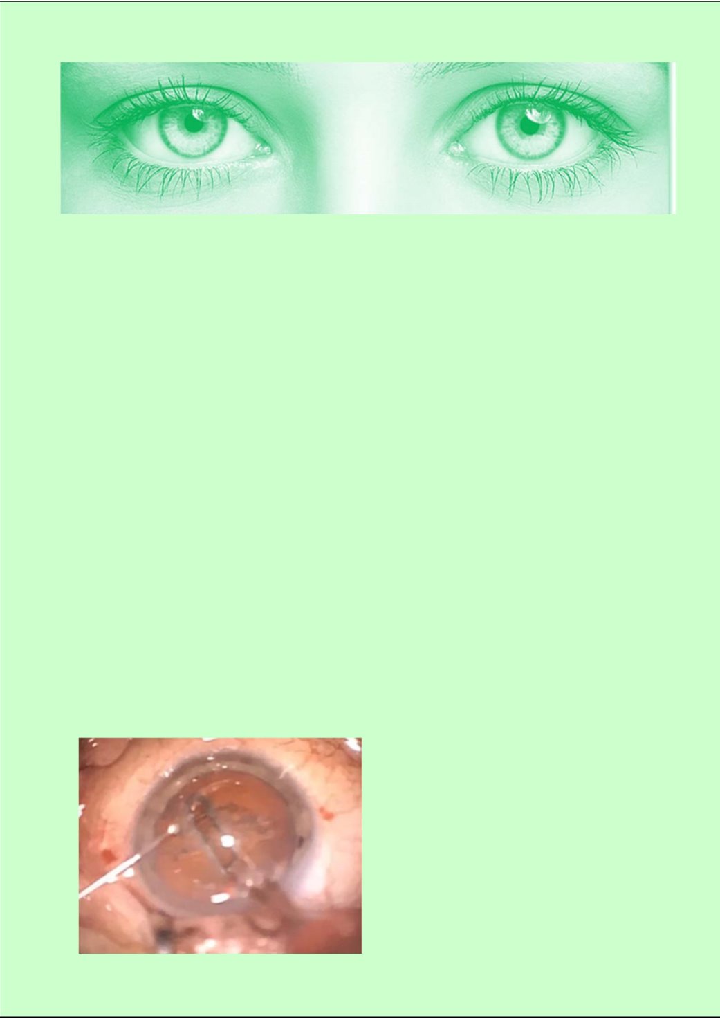
A photo that I took of a cataract surgery being
performed, specifically the lens capsule being
opened up.
Glaucoma:
The eye has a natural pressure inside.
When you have glaucoma the pressure is much
higher than normal and it damages the reƟna at
the back of the eye. To treat the problem the doc-
tors try to lower the pressure using drugs or if this
does not work they do surgery. The second opera-
Ɵon I watched was on a paƟent with glaucoma.
To lower the pressure the paƟent had a drain in-
serted into the front of the eye, the anterior
chamber, to allow the aqueous humor to drain out
faster than normal and reduce the internal pres-
sure. The drain had stopped working so the sur-
geon removed the drain and made a parƟal depth
hole in the side of the eye – Through the white
part – to drain the fluid and reduce the pressure.
The sclera has a lot of blood vessels in it and the
surgeon did use some Ɵny sutures.
AMD:
This disease is called Age-war elated Macu-
lar DegeneraƟon. The Macular is a part of the
reƟna and in this disease it gets damaged usu-
ally because cells start to grow in that area.
Ophthalmologists treat this problem by injecƟng
drugs into the vitreous – the jelly like substance
in the back of the eye – near the reƟna. These
drugs stop the cells growing and slow down the
damage.
he surgeon used a Ɵny needle and pushed it
through the sclera and got the end of the needle
close to the reƟna before injecƟng the drug. It on-
ly took a few minutes.