


 1469 / 1708

temporal lobes was 16 Gy with IMRT. This was reduced to 4
CGE with protons and 2 CGE with IMPT. A similar benefit
was seen with the dose received by the whole brain. Five
percent and 50% of the pituitary received 16 and 12 Gy
with IMRT, respectively. The dose to 5% and 50% of this
structure with both proton and IMPT plans was less than 1
CGE in each case. The hypothalamus received a mean dose
of 10.7 Gy with IMRT. For protons, mean dose was 0.2
CGE, and no measurable dose was delivered with IMPT.
Similarly, dose to the brainstem was reduced with proton
treatment. Dose–volume histograms
( Figs. 1 and 2 )visibly
show the benefit of protons for the brain and other CNS
structures.
Figure 4shows sagittal and coronal views and
illustrates the rapid dose falloff of proton radiation.
Similar to the infratentorial plan, greater sparing of CNS
structures was shown for proton and IMPT planning for the
supratentorial case. The hypothalamus was in close proximity
to the CTV for this particular case. The IMPT planning
provided substantially greater sparing for this particular
structure
( Fig. 5 ).
DISCUSSION
This study shows excellent early outcomes using proton
radiation for the treatment of patients with localized ependy-
moma. Consistent with several prior studies, we found
a significant correlation between subtotal resection and sub-
sequent local failure
(6, 28) .No significant late toxicity after
radiation was reported to date in patients followed up since
2000. Dose distributions for proton therapy compare
favorably with IMRT plans. The IMPT appears to allow for
further sparing of some critical structures.
Fortunately, disease control for childhood ependymoma
has improved significantly during the past several years,
and the 3- to 5-year survival rate range now is 60–80%
(7, 29–31). However, late side effects of radiation therapy
are still worrisome for this group of patients because of the
proximity of these tumors to critical tissues and the excep-
tionally young age at diagnosis.
Currently, the most widely available technique to mini-
mize toxicity to normal tissue without compromising dose
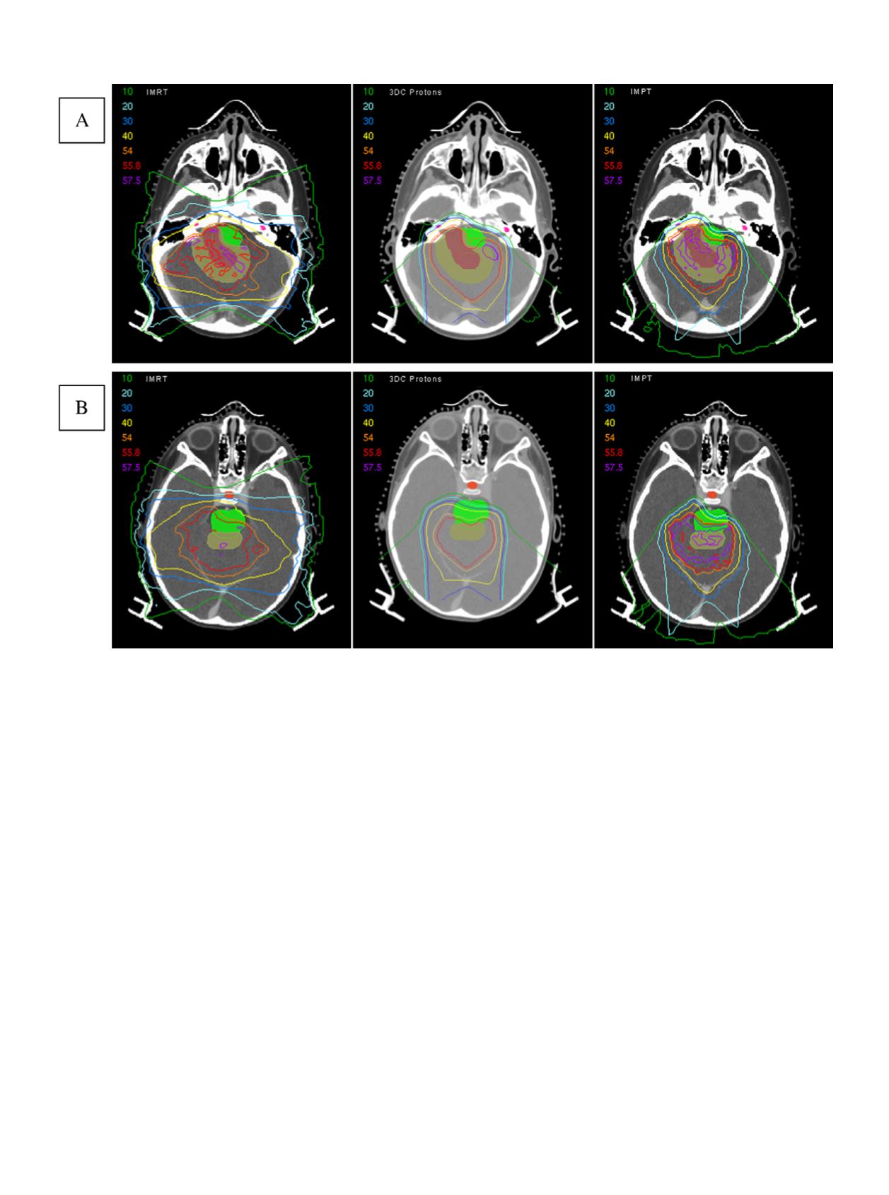
Fig. 3. Intensity-modulated radiation therapy (IMRT), proton, and intensity-modulated proton therapy (IMPT) plans
shown in the axial plane at the level of the (A) cochlea and (B) temporal lobes and pituitary gland. Gross tumor volume
(GTV) is shown in red, and clinical tumor volume (CTV) is shown in yellow. Protons show improved sparing of the
cochlea, cerebellum, pituitary gland, and temporal lobes. The IMPT plan shows superior proximal target conformity
and further sparing of structures.
Proton treatment of childhood ependymoma
d
S. M. M
AC
D
ONALD
et al
.
983