


 6 / 16

JOURNAL SCAN
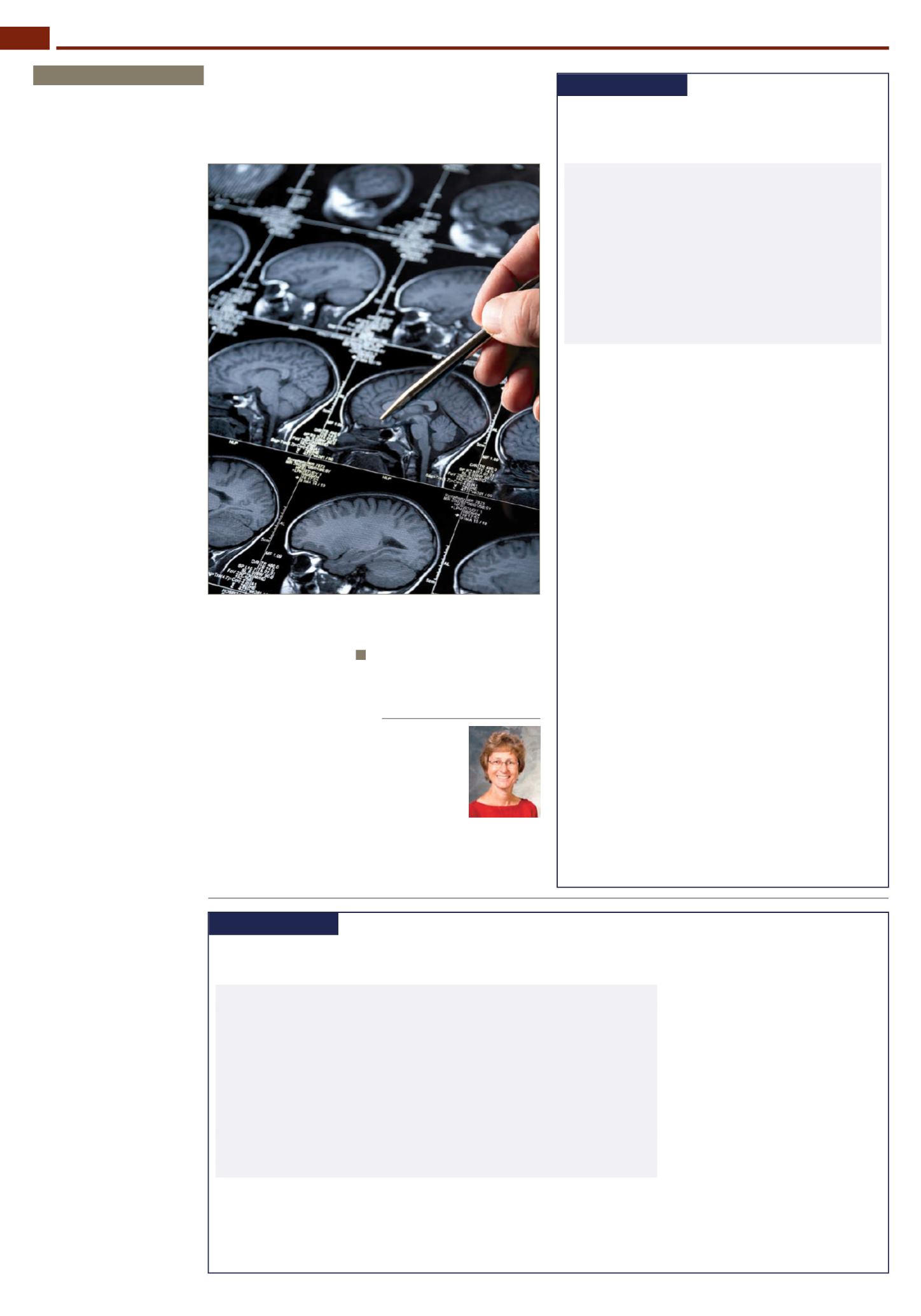
Anticholinergic use and cognition and brain
atrophy in cognitively normal older adults
JAMA Neurology
Take-home message
•
Well over 400 cognitively normal older adults were followed to evalu-
ate the association between use of anticholinergic (AC) medication and
cognitive impairment. People taking AC medication (AC+) showed lower
scores on immediate-recall memory testing, the Trail Making Test Part
B, and tests of executive function than people not taking AC medication
(AC−). In addition, neuroimaging revealed a reduction in total cortical
volume and temporal lobe cortical thickness and an increase in lateral
ventricle and inferior lateral ventricle volumes in AC+ individuals com-
pared with those who were AC−.
•
The use of AC medication should be avoided in older adults if a suitable
alternative is available due to the associated increase in brain atrophy
and clinical signs of cognitive decline.
Dr Irene Mace Hamrick
IMPORTANCE
The use of anticholinergic (AC) medication is linked to cognitive
impairment and an increased risk of dementia. To our knowledge, this is the first
study to investigate the association between AC medication use and neuroimag-
ing biomarkers of brain metabolism and atrophy as a proxy for understanding
the underlying biology of the clinical effects of AC medications.
OBJECTIVE
To assess the association between AC medication use and cognition,
glucose metabolism, and brain atrophy in cognitively normal older adults from
the Alzheimer’s Disease Neuroimaging Initiative (ADNI) and the Indiana Memory
and Aging Study (IMAS).
DESIGN, SETTING AND PARTICIPANTS
The ADNI and IMAS are longitudinal studies
with cognitive, neuroimaging, and other data collected at regular intervals in clini-
cal and academic research settings. For the participants in the ADNI, visits are
repeated 3, 6, and 12 months after the baseline visit and then annually. For the
participants in the IMAS, visits are repeated every 18 months after the baseline
visit (402 cognitively normal older adults in the ADNI and 49 cognitively normal
older adults in the IMAS were included in the present analysis). Participants were
either taking (hereafter referred to as the AC+ participants [52 from the ADNI
and 8 from the IMAS]) or not taking (hereafter referred to as the AC- participants
[350 from the ADNI and 41 from the IMAS]) at least 1 medication with medium or
high AC activity. Data analysis for this study was performed in November 2015.
MAIN OUTCOMES AND MEASURES
Cognitive scores, mean fludeoxyglucose F 18
standardised uptake value ratio (participants from the ADNI only), and brain
atrophy measures from structural magnetic resonance imaging were compared
between AC+ participants and AC- participants after adjusting for potential
confounders. The total AC burden score was calculated and was related to target
measures. The association of AC use and longitudinal clinical decline (mean
[SD] follow-up period, 32.1 [24.7] months [range, 6-108 months]) was examined
using Cox regression.
RESULTS
The 52 AC+ participants (mean [SD] age, 73.3 [6.6] years) from the
ADNI showed lower mean scores on Weschler Memory Scale-Revised Logical
Memory Immediate Recall (raw mean scores: 13.27 for AC+ participants and 14.16
for AC- participants; P=0.04) and the Trail Making Test Part B (raw mean scores:
97.85 seconds for AC+ participants and 82.61 seconds for AC- participants;
P=0.04) and a lower executive function composite score (raw mean scores:
0.58 for AC+ participants and 0.78 for AC- participants; P=0.04) than the 350
AC- participants (mean [SD] age, 73.3 [5.8] years) from the ADNI. Reduced total
cortical volume and temporal lobe cortical thickness and greater lateral ventricle
and inferior lateral ventricle volumes were seen in the AC+ participants relative
to the AC- participants.
CONCLUSIONS AND RELEVANCE
The use of AC medication was associated with
increased brain atrophy and dysfunction and clinical decline. Thus, use of AC
medication among older adults should likely be discouraged if alternative thera-
pies are available.
Association between anticholinergic medication use and cognition, brain me-
tabolism, and brain atrophy in cognitively normal older adults
JAMA Neurol
2016 April 18 [EPub ahead of print]. S Risacher, B McDonald, E Tallman, et al.
EXPERT OPINION
Anticholinergic meds and
cognition
By Dr Irene Mace Hamrick
A
longitudinal study published
last month in
JAMA Neurol-
ogy
collected neuroimaging
and cognitive testing results of 451
cognitively intact adults at specific
intervals over 32.1 months (range
6–108 months).
1
The mean age of
study participants was 73.3 years,
and 60 participants had been taking
medium- or high-potency anticholin-
ergic medications for a minimum of
1 month. Over the follow-up period,
the group exposed to anticholinergic
medications had worse outcomes on
Wechsler Memory Scale, Trail Mak-
ing Test Part B, and lower glucose
metabolism on PET. MRI showed
reduction of brain volume overall
and in the hippocampus, along with
enlargement of ventricles.
Alzheimer’s disease is considered
to be a deficit of acetylcholine
(ACh), and most medications for
the treatment enhance ACh in the
brain by inhibiting the enzyme that
breaks it down. In contrast, medi-
cations that inhibit ACh should not
be used in older adults, especially
those at risk or with the diagnosis
of dementia. Since 1992, the Beers
Criteria recommend against the
use of anticholinergic medications
in older adults.
2,3
More data are
emerging that changes associated
with anticholinergic medications are
permanent, with one study last year
showing a 56% increase of demen-
tia and 68% increase of Alzheimer’s
disease with >1095 cumulative,
standardised daily doses.
4
With the new prospective, ad-
ditional findings on brain imaging
and cognitive testing, what are we
to do when patients come to us with
insomnia or incontinence that we
often treat with anticholinergics?
Anticholinergics, such as diphen-
hydramine, which is in all “PM”
over-the-counter products, lose their
sleep-inducing efficacy after only 3
nights.
5
Explaining to our patients
that sleep needs decrease and sleep
gets more fragmented with increas-
ing age can reassure many worries.
Lowering the temperature of the
bedroom and moving bath time to
earlier in the evening gives the brain
the signal that it is time to go to
sleep as the body temperature drops.
Almost all medications on the
market for urge incontinence are
anticholinergic, and, yes, antimus-
carinics have the same effect on the
brain. Kegel exercises, if done often
(>45 times daily), are as effective as
oxybutynin, even in men.
6
Avoiding
bladder irritants such as alcohol,
caffeine, and nicotine can prevent
incontinence. Many patients restrict
their fluid intake in an attempt to
avoid bladder accidents or to save
trips to the bathroom, and do not
feel thirsty due to apoptosis of the
thirst centre in the hypothalamus. A
concentrated urine is very irritating
to the bladder, and increasing fluid
intake to at least 2 quarts a day can
prevent urge incontinence. Extra ef-
forts on our part can have significant
benefits for our patients’ brains.
References
1. Risacher SL, McDonald BC, Tallman EF.
Association between anticholinergic
medication use and cognition, brain
metabolism, and brain atrophy in cogni-
tively normal older adults.
JAMA Neurol
[Published online April 18, 2016]
2. Beers MH, Ouslander JG, Fingold SF, et
al. Inappropriate medication prescribing
in skilled-nursing facilities.
Ann Intern
Med
1992;117(8):684–689.
3. American Geriatrics Society 2015 Updat-
ed Beers Criteria for Potentially Inappro-
priate Medication Use in Older Adults.
J
Am Geriatr Soc
2015;63(11):2227–2246.
4. Gray SL, Anderson ML, Dublin S, et al.
Cumulative use of strong anticholiner-
gics and incident dementia: a prospec-
tive cohort study.
JAMA Intern Med
2015;175(3):401–407.
5. Richardson GS, Roehrs TA, Rosenthal
L, et al. Tolerance to daytime sedative
effects of H1 antihistamines.
J Clin Psy-
chopharmacol
2002;22(5):511–515.
6. Burgio KL, Goode PS, Johnson TM, et
al. Behavioral versus drug treatment
for overactive bladder in men: the
Male Overactive Bladder Treatment in
Veterans (MOTIVE) Trial.
J Am Geriatr
Soc
2011;59(12):2209–2216.
Irene Mace
Hamrick MD
FAAFP, AGSF
is an associate
professor, Clinical
Health Sciences,
Department of
Family Medicine,
University of
Wisconsin; director of geriatrics
services, Department of
Family Medicine, University of
Wisconsin, Madison, Wisconsin
JOURNAL SCAN
Brain stimulation and constraint for perinatal stroke hemiparesis
Neurology
Take-home message
•
Effective treatments for cerebral palsy (CP) are limited. Hemiparetic CP is a common type, and often results
from in-utero or perinatal stroke. Constraint-induced movement therapy (CIMT) and repetitive transcranial
magnetic stimulation (rTMS) are therapeutic options based on engaging neuroplasticity. This study is a
2x2 factorial design, randomised, blinded controlled trial in which hemiparetic children aged 6 to 19 years
underwent a 2-week goal-directed training camp during which they had daily CIMT, rTMS, both, or neither.
Using the Assisting Hand Assessment (AHA) and the Canadian Occupational Performance Measure as
primary outcomes, all interventions showed benefit at 1 week to 2 months after the intervention, with
some of the benefit waning by 6 months. The addition of rTMS, CIMT, or both doubled the chances of
clinically significant improvement, and the effects were additive. The largest improvement was seen with
the combined rTMS+CIMT modality, with an improvement of 5.91 AHA units, compared with 0.62 units
in patients receiving neither. The therapies were well-tolerated and all participants completed the trial.
•
The results show a role for the combined rTMS and CIMT approach for improving therapy-induced
functional motor gains in hemiparetic CP.
Dr Codrin Lungu
OBJECTIVE
To determine whether the addition of re-
petitive transcranial magnetic stimulation (rTMS) and/
or constraint-induced movement therapy (CIMT) to
intensive therapy increases motor function in children
with perinatal stroke and hemiparesis.
METHODS
A factorial-design, blinded, randomised con-
trolled trial (clinicaltrials.gov/NCT01189058) assessed
rTMS and CIMT effects in hemiparetic children (aged
6-19 years) with MRI-confirmed perinatal stroke. All
completed a 2-week, goal-directed, peer-supported
motor learning camp randomised to daily rTMS, CIMT,
both, or neither. Primary outcomes were the Assisting
Hand Assessment and the Canadian Occupational
Performance Measure at baseline, and 1 week, 2 and
6 months postintervention. Outcome assessors were
blinded to treatment. Interim safety analyses occurred
after 12 and 24 participants. Intention-to-treat analysis
examined treatment effects over time (linear mixed ef-
fects model).
RESULTS
All 45 participants completed the trial. Addition
of rTMS, CIMT, or both doubled the chances of clinically
significant improvement. Assisting Hand Assessment
gains at 6 months were additive and largest with rTMS
+ CIMT (β coefficient = 5.54 [2.57-8.51], p = 0.0004). The
camp alone produced large improvements in Canadian
Occupational Performance Measure scores, maximal
at 6 months (Cohen d = 1.6, p = 0.002). Quality-of-life
scores improved. Interventions were well tolerated
and safe with no decrease in function of either hand.
CONCLUSIONS
Hemiparetic children participating in
intensive, psychosocial rehabilitation programs can
achieve sustained functional gains. Addition of CIMT
and rTMS increases the chances of improvement.
CLASSIFICATION OF EVIDENCE
This study provides Class
II evidence that combined rTMS and CIMT enhance
therapy-induced functional motor gains in children with
stroke-induced hemiparetic cerebral palsy.
Brain Stimulation and Constraint for Perinatal
Stroke Hemiparesis: The PLASTIC CHAMPS Trial
Neurology
2016 May 03;86(18)1659-1667, A Kirton,
J Andersen, M Herrero, A Nettel-Aguirre, L Carsolio,
O Damji, J Keess, A Mineyko, J Hodge, MD Hill
GENERAL NEUROLOGY
PRACTICEUPDATE NEUROLOGY
6