


 10 / 28

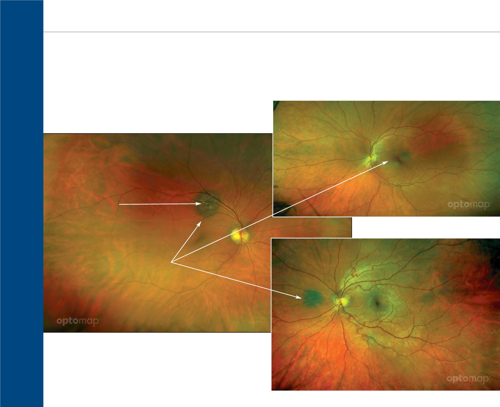
Choroidal Nevi
8
Choroidal Nevi
are a flat, benign pigmented area that appears in the back of the eye. Studies have shown the benefit
of imaging choroidal nevi using a widefield scanning laser ophthalmoscope in that the two imaging
channels ( red 633nm and green 532nm) can be used to help determine the presence of choroidal
nevi. Utilizing the ultra-widefield SLO increased the prevalence of visualizing choroidal nevi compared
to other population-based studies where an
ultra-widefield SLO was not used.
1
Choroidal Nevi
Drusen in the nevus
1. Gordon-S haag A , Barnard S , Millodot M, Gantz L, Chiche G, Vanessa E , R uth W, Pinchasov R , Gosman
Z, S imchi M, Koslowe K & S hneor E . Prevalence of choroidal naevi using scanning laser ophthalmoscope.
Ophthalmic Physiol Opt 2014, 34, 94–101.