


 952-953 / 3107


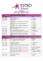
Clinical example: Prostate XRT
Transverse and Sagital kV cone beam CT images with bladder (green),
prostate (yellow), PTV (blue) and rectal contours (red) superimposed. Can be
difficult to identify edge of prostate but rectal-prostate interface is easily seen