


 26 / 140


24
Chapter 4
Primary Care Otolaryngology
Foreign Bodies
Foreign bodies can present as airway emergencies. Usually, however, by
the time the patient gets to the emergency room, the foreign body in the
airway has been expelled (often by the
Heimlich maneuver
), or else the
patient is no longer able to be resusci-
tated. Foreign bodies in the
pharynx
or
laryngeal inlet
can often be
extracted by
Magill forceps
after
laryngeal exposure with a standard
laryngoscope. The patient will usually
vomit, so
suction is mandatory.
Bronchial foreign bodies
will require
operative
bronchoscopy
for removal.
Occasionally, a tracheotomy will be
required, such as for a patient who has
aspirated a partial denture with
imbedded hooks. Children often aspi-
rate peanuts, small toys, etc., into their
bronchi. Occasionally these patients
present as airway emergencies,
although they more typically present
with
unexplained cough or pneumo-
nia
. Chevalier Jackson, the famous
bronchoscopist
, has noted, “All that
wheezes is not asthma.” In other
words, always remember to think of
foreign body aspiration when a pedi-
atric patient presents with unexplained cough or pneumonia. If a
ball-
valve obstruction
results,
hyperinflation of the obstructed lobe or seg-
ment
can occur. This is easier to visualize on
inspiration-expiration
films
.
Mucormycosis
This is a
fungal infection
of
the sinonasal cavity that occurs in
immuno-
compromised
hosts. Typically it appears in patients receiving bone mar-
row transplantation or chemotherapy. It is a devastating disease, with a
significant associated mortality.
Mucor
is a ubiquitous fungus that can
become
invasive
in susceptible patients, classically those with diabetes
with
poor glucose regulation
who became
acidotic
. If there is any other
system failure
(e.g.,
renal failure
), mortality goes up significantly. The
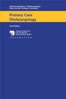
Figure 4.3.
A coin is seen here trapped in the patient’s
esophagus.