


 2 / 28

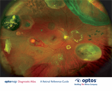
Optos’ core devices produce ultra-widefield (UWF™), high resolution
digital images (
opto
map
®
) of approximately 82% and 200° of the retina,
something no other device is capable of doing in any single image.
An
opto
map image provides a bigger picture and more clinical information
which facilitates the early detection, management and effective treatment of
disorders and diseases evidenced in the retina such as retinal detachments
and tears, glaucoma, diabetic retinopathy and age-related macular
degeneration. Retinal imaging can also indicate evidence of non-eye or
systemic diseases such as hypertension and certain cancers.
opto
map images consist of two channels of information, a red channel
(633nm) which visualises the choroidal layer and a green channel (532nm)
which visualises the retinal pigment epithelium.
The
opto
map Diagnostic Atlas:
A Retinal Reference Guide
is designed to
illustrate how different pathologies are visualised on ultra-widefield images.
Reference for Definitions
Dictionary of Eye Terminology. Sixth Edition. 2012.
Barbara Cassin and Melvin L. Rubin, MD.
Triad Communications, Inc.
Diagnostic Atlas
A Retinal Reference Guide