


 8 / 16

8
We live in a three-dimensional world, yet most physicians and
surgeons diagnose, treat and operate on their patients using flat
images that often present a less-than-perfect reflection of what is
going on inside the body. But as with so many aspects of daily
life, cutting-edge technology is providing a decisive upgrade to
that process.
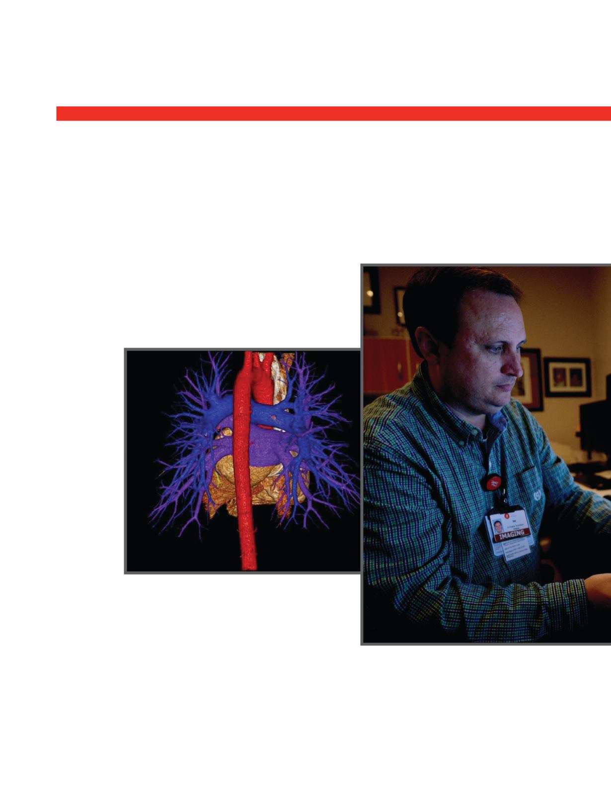
Increasingly, 3-D imaging is providing a clearer, less invasive
and more realistic view of joints and organs that is proving
to be especially valuable in pediatrics. That’s why Children’s
of Alabama established one of the first pediatric 3-D imaging
labs when its 12-story, 760,000-square-feet expansion opened
in 2012. Jon Betts, RT(R)(MR), serves as a 3-D Imaging
Technologist in the lab, working closely with physicians in a
number of
services
to provide
the post-
processing
images they
need to form
effective
treatment
plans. Data
is taken
from regular
scans and
reformatted
using various
software
platforms to
produce the
3-D image.
“I ask them,
‘What are
your needs?’
And then I figure out how to give them what they need, using
several different systems,” Betts said. “The scans are done with
standardized data sets. They can then be layered to stack as
many data sets as needed to create the 3-D image.”
At Children’s, 3-D imaging is playing a vital role in surgery.
“It helps the surgeon visualize the procedure and make the
best plan,” Betts said. “It also helps patients and their parents
understand what will be done during the surgery.”
In Pediatric Neurosurgery, the 3-D images are especially
effective for treating epilepsy. Scans are made during seizures
to find the focus of the epilepsy and used to help plan the
procedure. They are also used in conjunction with a surgical
guidance computer in the OR during the procedure to pinpoint
the area of concern.
Orthopedic surgeon Michael Conklin, M.D., has found these
enhanced views, which can be manipulated and rotated on the
computer screen to provide a 360-degree perspective, to be
invaluable when evaluating particularly complicated structural
anomalies such as kyphoscoliosis and planning surgical
correction of such deformities. “It’s difficult to appreciate the
deformity when looking at just one plane,” he said. “Three-D
imaging allows us to look at each vertebra.”
Conklin and his colleagues also use the technology to monitor
post-surgical healing of fractures, particularly those that require
screws, pins and other hardware. The 3-D image shows in great
Lab Calls on 3-D Imaging
To Treat Patients
Inside Technology