


 601 / 1116

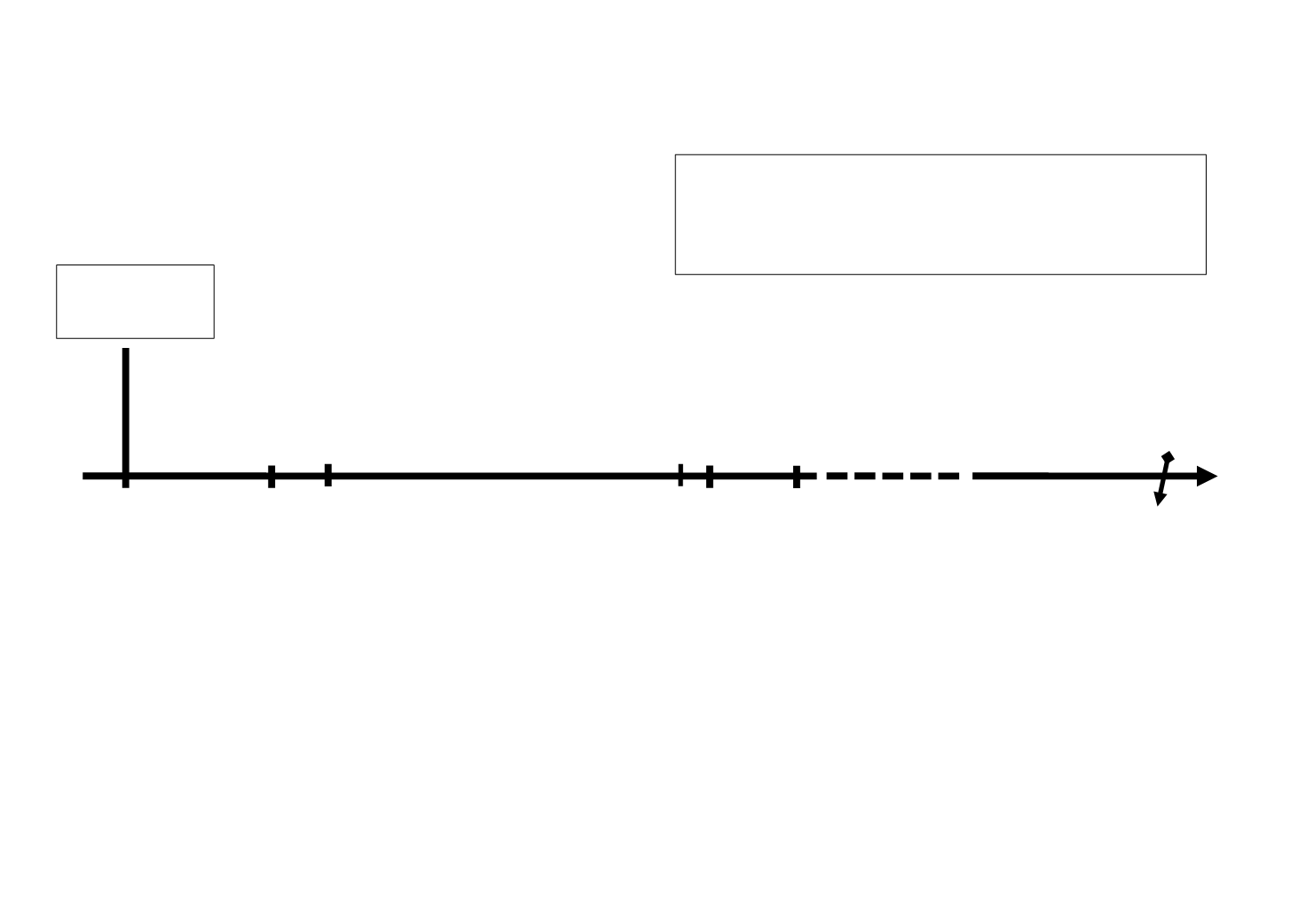
3 weeks
MRI
P
atient
inclusion
staging
Patient follow-up - 2 years: clinical/CT
Remission
continue follow-up
Lesion progression
biopsy (+)
treatment
biopsy (-)
continue follow-up
Correlation imaging-outcome
2 years endpoint
Start
CRT
End
CRT
Baseline
MRI
Lesion
Identification
Routine imaging
3 Months CT