


 148 / 242


Chapter 6: Temporal Bone Fractures
Resident Manual of Trauma to the Face, Head, and Neck
146
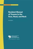
Figure 6.4
Axial view of the right
temporal bone with a
transverse fracture (red
dashed line) crossing
the petrous bone and
involving the lateral
aspect of the IAC.
2. Transverse Fractures
Transverse fractures cross the petrous ridge and have a higher inci-
dence of otic capsule involvement. These fractures require more energy
and classically result from a blow to the occipital region. Transverse
fractures are more often associated with inner ear injury, resulting in
SNHL, and have a higher incidence of facial nerve injury. Figure 6.4
demonstrates the radiologic appearance of a transverse fracture. This
patient sustained his fracture in a motor vehicle accident and had
normal facial nerve function but lost all hearing. Although this system is
simple and easy to understand, many fractures have mixed patterns,
limiting this system’s utility.
B. Otic Capsule-Sparing versus Otic Capsule-Involving
Classification
This classification system is based on the presence or absence of
involvement of the otic capsule. This system was introduced to empha-
size the functional sequelae of the fracture. Results from the two series
proposing this classification scheme indicate that 2.5–5.8 percent of
fractures involve the otic capsule. Figure 6.3 illustrates an otic capsule-
sparing fracture, while figure 6.4 illustrates an otic capsule-involving
fracture.