


 59 / 242

 www.entnet.org
57
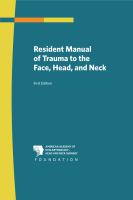
Figure 3.19
Type I medial canthal
region fracture, unilateral.
Figure 3.20
Type II medial canthal
region comminuted
fracture, right medial
orbit.
drainage system will produce epiphora, which requires further evalua-
tion with lacrimal probing at the time of the surgical repair.
C. Classification of Medial Canthal Tendon Injuries
Medial canthal tendon injuries are classified according to three types:
y
y
Type I:
Single-fragment bone segment with intact canthal tendon
insertions (Figure 3.19).
y
y
Type II:
Comminuted central bone segment with fractures remaining
external to the medial canthal tendon insertion (Figure 3.20).
y
y
Type III:
Comminuted single fragment with fractures extending into
bone bearing the canthal insertion.
The injury type can be identified on imaging studies and confirmed at
surgical exploration and repair.
Though uncommon, blunt trauma can also result in a rupture of the
anterior and/or posterior insertions of the canthal slips near their
insertion on the lacrimal fossa. It would be seen in the lax eyelid,
without evidence of distracted lacrimal fossa fractures.
D. Lacrimal System Injuries
Laceration of the medial eyelids with discontinuity of the lacrimal
canaliculi will be seen in vertical lacerations medial to the puncta. Deep