


 11 / 24

THE GEC ESTRO HANDBOOK OF BRACHYTHERAPY | Part II: Clinical Practice
Version 1 - 25/04/2016
Endometrial Cancer
11
8.2.2 Technique of application
The easiest way to perform such an application is under spinal
or general anaesthesia, but it may be performed using a combi-
nation of systemic and local analgesia with or without sedation.
The patient is positioned in the dorsal lithotomy position and
a bladder catheter inserted. The procedure starts with a clinical
examination including abdominal and rectovaginal bimanual in-
vestigation in order to confirm the pathologic anatomy and the
position and size of the uterus.
Transabdominal ultrasound at this time is also very valuable to
confirm the relation between the tube and the uterine cavity.
Depending on the technique of application, variable dilatation of
the cervical os and canal is indicated increasing with the number
of catheters to be introduced. The number of catheters depends
on the individual dimensions of the uterine cavity.
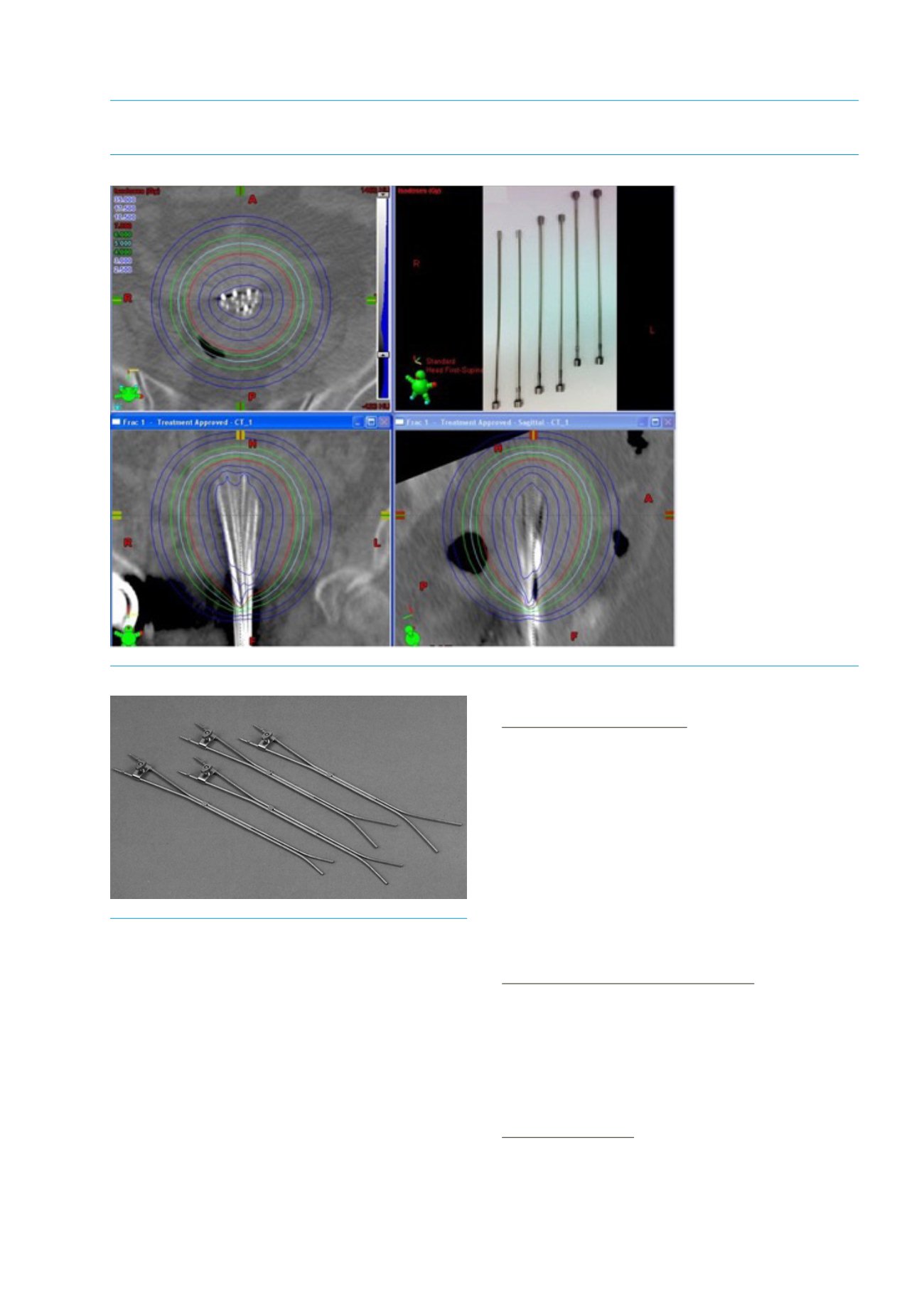
Norman Simon capsule packing.
A large uterine cavity being treated with Norman-Simon cap-
sules (modified Heyman’s capsules) will usually need more than
10 catheters, which requires as wide a dilatation as possible (up
to Hegar 10 - 12). The packing is complete after the uterine cavity
has been filled, but is usually extended to the uterine cervix. A
capsule in the cervical canal will prevent this closing down dur-
ing treatment and make removal easier. This is of particular im-
portance in PDR treatment with a longer time period of many
hours or days, as the internal cervical os may become narrow
again and prevent extraction of the tubes with the capsules. The
number of tubes applied varies significantly with the individual
anatomy, but between 5 and 18 is typical. Finally, the vagina is
packed or a mould is introduced, to keep the applicators in place.
Two or three channel-applicators (Y-shaped)
One of the two curved Rotte applicators is introduced and the
end is gently advanced towards one corner of the uterine fundus
taking into account the measured length of the uterine cavity.
The second one is introduced in the same way into the oppo-
site corner. Both applicators are finally fixed together by a screw
clamp on the applicator stem. The whole applicator is stabilized
by vaginal packing.
One channel-applicator
The intrauterine tube is introduced into the uterine cavity as far
as the measured intrauterine length. This length is defined in ad-
vance by a flange on the metallic tube so that the applicator is
fixed in front of the cervical os. The vaginal fixation is achieved
with a cylindrical applicator advanced over the metallic tube and
pressed against the flange.
Figure 15.5: Norman Simon capsules (top right) and CT planning images of capsules in situ with CTV and isodoses
Figure 15.6: Rotte Y applicators