


 6 / 24

THE GEC ESTRO HANDBOOK OF BRACHYTHERAPY | Part II: Clinical Practice
Version 1 - 25/04/2016
Endometrial Cancer
6
ence of 2 of the following: age >60yrs, Grade 3, >50%myometrial
invasion or LVSI.
An alternative classification divides endometrial cancers into
Type I and Type II. Type I is characterised by women who are
obese, have hyperlipidaemia, signs of hyperoestrogenisation,
anovulatory uterine bleeding, late onset of menopause and in-
fertility with hyperplasia of the ovaries and endometrium. These
features are associated with grade 1 or 2 cancers, superficial inva-
sion and high progesterone sensitivity with a relatively favoura-
ble outcome having a 78% 5 year survival compared to only 59%
in those cases without these features [17].
A more sophisticated analysis from which a nomogram to indi-
vidualise risk based on age, grade, myometrial invasion and LVSI
has been constructed for locoregional relapse, disease free and
overall survival. [17].
5. WORK UP
In approximately 5-10% of patients presenting with post-menopausal vaginal bleeding, endometrial cancer is the under-
lying cause. Systematic work-up includes the following: history,
general and gynaecological examination and transvaginal ultra-
sound which provides information on endometrial thickness
and tumour extent as shown in figure 15.2 followed by pipelle
sampling, EUA for systematic biopsies of both endometrium
and cervix with hysteroscopy or fractional curettage. Ultrasound
may also be useful both in screening high risk patients such as
those on prolonged tamoxifen and in evaluating premenopausal
women. Cystoscopy and rectoscopy are indicated in advanced
disease.
For the majority of stage I low grade patients a chest radiograph
and transvaginal ultrasound combined with gynaecological
examination are sufficient to assess the disease extent prior to
surgery. For patients with more advanced stage disease and high
grade histology, CT of the chest and abdomen is used to screen
for involved lymph nodes and rule out distant metastasis, while
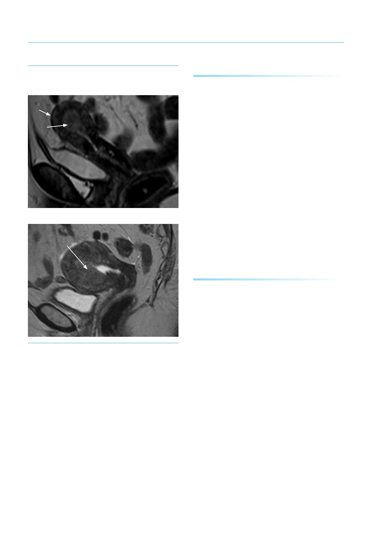
pelvic MRI is recommended for evaluating the local tumor ex-
tent. MRI will correctly predict the surgical stage in 70-80% of
cases [19] as shown in figure 15.3.
6.
INDICATIONS FOR BRACHYTHERAPY
Surgery consisting of hysterectomy and bilateral oophorectomy
is the most important treatment for the majority of endometrial
cancer patients. More than half of the patients will not require any
further adjuvant treatment and have an excellent 95% recurrence
free survival. The most frequent indication for brachytherapy is
that of postoperative treatment, where the aim is to prevent local
vaginal recurrence. Less frequent indications for brachytherapy
are the primary treatment in patients that are no surgical candi-
dates and the treatment of recurrent vaginal disease.
6.1 Postoperative radiotherapy
With the advent of risk based adjuvant radiotherapy, it was
already recognised that patients with grade 1 and 2 endo
metrioid type tumours without invasion in the myometrium had
a very low risk of disease recurrence with surgery alone. The role
of post-operative radiotherapy for stage I intermediate risk en-
dometrial cancer has been subject to a number of multicentre
randomised trials in recent years focussing in all but one case
principally on the use of external beam irradiation.
The PORTEC 1 study [20] randomised 714 stage I intermediate
risk patients with at least one risk feature on histology,>50%
myometrial invasion, grade 2 or grade 3 (excluding >50% in-
vasion AND grade 3) to receive either post-operative external
beam treatment delivering 46Gy in 23 fractions or no adjuvant
postoperative radiotherapy. Mature results have confirmed a 9%
reduction in pelvic relapse (5% with postoperative radiotherapy
versus 14% without at 5-years) in this patient population but no
reduction in distant metastasis or in endometrial cancer deaths.
The GOG-99 study [21] included 392 surgically staged patients
and was similar in design but included lymphovascular invasion
Figure 15.3: Diagnostic MRI scan to show localized uterine cancer (a) not invading the myome-
trium and (b) invading >50% of myometrium: Stage IB
a. Non-muscle invasive endometrial cancer (IA)
b. Stage IB showing extensive invasion of uterine wall
Intact wall
of uterus
Tumour
Extensive tumour invading
and distorting muscle wall;
compare with intact muscle
wall above in 15.3.(a)