


 7 / 56

The Cells of the Heart
15
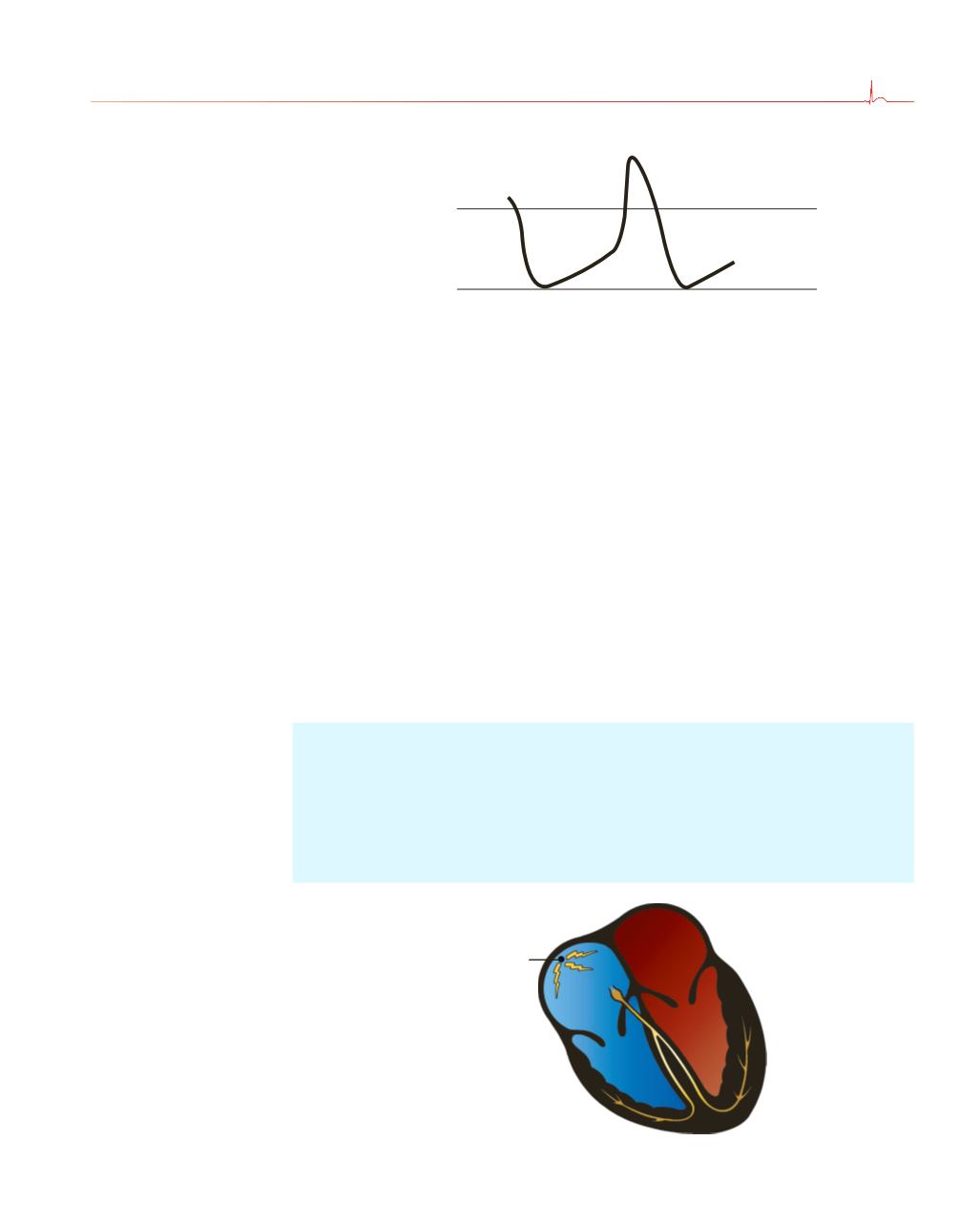
The dominant pacemaker cells in the heart are located high up
in the right atrium. This group of cells is called the
sinoatrial (SA)
node
, or
sinus node
for short. These cells typically fire at a rate of
60 to 100 times per minute, but the rate can vary tremendously
depending upon the activity of the autonomic nervous system (
e.g
.,
sympathetic stimulation from catecholamines, such as epinephrine and
norepinephrine, accelerates the sinus node, whereas vagal stimulation
slows it) and the demands of the body for increased cardiac output
(exercise raises the heart rate, whereas a restful afternoon nap lowers it).
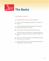
0 mV
−
60 mV
A
B
C
D
The electrical depolarization–repolarization cycle of a cardiac pacemaker
cell. Point
A
is the minimal negative potential. The gentle rising slope
between points
A
and
B
represents a slow, gradual depolarization. At
point
B
, the threshold is crossed and the cell dramatically depolarizes
(as seen between points
B
and
C
); that is, an action potential is produced.
The downslope between points
C
and
D
represents repolarization. This
cycle will repeat over and over for, let us hope, many, many years.
Pacemaker cells are really good at what they do. They will continue firing
in a donor heart even after it has been harvested for transplant and before
it has been connected to the new recipient. The transplanted heart, devoid
of normal vagal stimulation (the nerves are cut when the new heart is
implanted), beats at an average rate of 100 beats per minute (bpm).
Sinus node
In a resting individual, the sinus node typically fires 60 to 100 times per
minute, producing a regular series of action potentials, each of which
initiates a wave of depolarization that will spread through the heart.