


 2 / 28

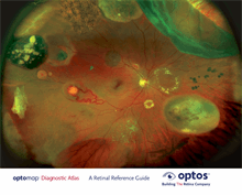
Optos' core devices produce ultra-widefield (UWF™ ) , high resolution
digital images (
opto
map
®
) of approximately 82% and 200° of the retina,
something no other device is capable of doing in any single image.
An
opto
map image provides a bigger picture and more clinical information
which facilitates the early detection, management and e ective treatment of
disorders and diseases evidenced in the retina such as retinal detachments
and tears, glaucoma, diabetic retinopathy and age-related macular
degeneration. Retinal imaging can also indicate evidence of non-eye or
systemic diseases such as hypertension and certain cancers.
opto
map images consist of two channels of information, a red channel
(633nm) which visualizes the choroidal layer and a green channel (532nm)
which visualizes the retinal pigment epithelium.
The
opto
map Diagnostic Atlas:
A Retinal Reference Guide
is designed to
illustrate how di erent pathologies are visualized on ultra-widefield images.
Reference for Definitions
Dictionary of Eye Terminology. Sixth Edition. 2012.
Barbara Cassin and Melvin L. Rubin, MD.
Triad Communications, Inc.
Diagnostic Atlas