


 8 / 52

C
ancer
T
reatment
<<< 5
6
Mei/May 2015
vet
nuus
•
news
skin to a depth of 4/5 cm. Practically
this means that tumours that you can
see and feel can be treated with elec
trons. The electrons suffice for cutane
ous tumours and tumours found in the
muscles of the limbs and trunk. Primary
tumours that might metastasise are
usually treated together with the clos
est lymph node as a prophylactic
measure. Photons are usually used for
human cancers and can penetrate very
deeply into the body. Very expensive
imaging is necessary for photon radia
tion therapy and a dedicated animal
linear accelerator machine would be
necessary if internal organs were to be
treated with photons, consequently this
kind of radiation is rarely used in South
Africa by veterinarians.
Before choosing electron radiation
as treatment the following facts must
be considered; the type of cancer, its
sensitivity to radiation, location, size,
stage, prognosis and cost. Dividing
cancer cells are more sensitive to
radiation than normal dividing cells.
The treatment is therefore designed to
kill off the cancer cells while giving a
reasonable number of healthy cells a
chance to survive and recover.
Different kinds of cancers have diffe
rent sensitivities to radiation, the broad
categories are determined by the rate
of their cells’ normal life cycle.
• High:
Cells of haemopoietic
(Mast Cell Tumour)
and lymphoid origin.
• Moderate:
Cells of epithelial
origin (carcinomas).
• Low: Cells of
mesenchymal origin
(sarcomas).
Fractionated low dose radiation is
extremely important for tumour
consolidation. The damaged DNA
that you do not hit today hopefully
will be undergoing mitosis at the next
treatment.
The clinical application of
radiation
During the last fourteen years I have
treated more than 1500 clinical cases
referred by veterinarians for radiation
therapy. Some of these cases have
been treated pre-operation, others
post-operation and some both pre-
and post-surgery. There is a 50/50
ratio dogs to cats.
The clinical cases recorded in this
study of dogs show:
• 50% Squamous cell carcinoma
(SCC)
• 26% Mesenchymal Cell Tumour
(sarcomas)
• 24% Mast Cell Tumour (MCT)
The clinical cases recorded in this
study of cats show:
• 95% SCC
• 5% “others”.
These figures may serve as useful
pointers to other veterinarians.
Although there is no pain associated
with electron radiation, the animals
do have to be immobilised for the
duration of the treatment (1-2minutes),
after which the sedation is reversed.
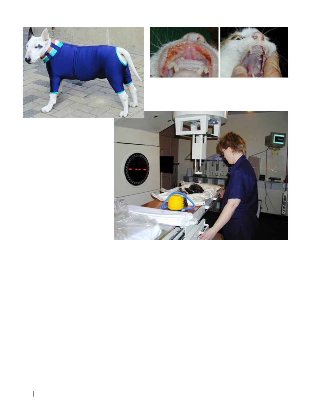
Linear accelerator radiation unit.
UV-Blocking body suit
Eosinophilic Granuloma
On 13 Apr 2005 this patient was in extreme pain and unable to eat or
groom. The second photograph shows the same patient seven months
later (24 Nov 2005). (The cat died 43 months later from a non-related
disease; the lesion did not recur)
Lead Article
I Hoofartikel
>>> 7