


 9 / 52

Mei/May 2015
7
vet
nuus
•
news
Squamous cell carcinomas
(SCC) in dogs and cats
This cancer is caused by high levels
of exposure to the B-fraction of ultra-
violet radiation (UVB), on susceptible
non-pigmented animal skin. South
Africa has one the highest UV levels
recorded in the world. Factors that
influence UVR levels across the globe
include altitude, intensity and duration
of sunlight, and the thickness of the
ozone layer. The UVB energy enters
the basal cell of the epidermis and
mutates the DNA, which results in
the uncontrolled proliferation of the
squamous cells. The normal histology
of the well-ordered squamous cell
tissue changes from its regular pattern
to one of disintegration, ultimately
leading to the development of a SCC.
Typically the clinical lesions are
treated with linear accelerator electron
radiation therapy with/without surgery.
The earlier this condition is recognised
and treated the better the prognosis.
To prevent the solar exposure in dogs,
ultra violet Lycra body suits are worn
(9 different
sizes and
male/female
are available)
together
with a long
acting 8 hour
sunscreen.
Cats are
naturally
nocturnal
animals, so the
cats that are at
risk should be
confined to the
sunniest room
in the house, with a special plastic
film that blocks the UV, but allows the
visible light and infrared energy to
pass through. The cats are fed in this
room at 08:00 and released at 17:00.
Cutaneous Mast Cell Tumours
(MCT)
MCT are visibly the most difficult
tumours to judge and a fine needle
aspirate of any suspicious “lump”
should be examined; confirmation
using a biopsy may be needed .
The pathology report is useful for
understanding how aggressive the
tumour is, by its mitotic index and
the description of the cells. Often the
pathologists do not grade the tumour,
as the prognosis and the eventual
outcome are vastly different. Radiation
for the primary cutaneous tumour is a
most effective way to consolidate the
tumour before surgery. If the tumour
has metastasized (detected by use of
ultrasound examination) both radiation
for the primary and chemotherapy for
the secondary tumours is necessary.
The MCT
patient
shown in
the photo
graphs was
graded as
Grade 3 and
yet did not
metastasise;
some of the
Grade 2 MCT
did meta
stasise.
Soft tissue sarcomas
are derived from the
mesenchymal cells
There are many tumours in
this category, e.g. liposarcoma,
chondrosarcoma, fibrosarcoma ,
peripheral nerve sheath tumours
and more. Biologically they generally
behave in a similar way; usually
they are found in the subcutaneous
tissue, they grow slowly, usually
do not metastasise, but often
recur after surgical excision. These
tumours require very wide surgical
margins as they are surrounded by a
pseudocapsule that allows the cancer
cells to escape. If they are radiated
pre-operation (consolidation), they
are easier to remove in their entirety.
It is easy to make the radiation field
very much larger than the surgical
field and if the area is also radiated
post-surgery, the success of the
treatment is greatly enhanced.
Because these tumours grow more
slowly than carcinomas, they require
a higher total dose of radiation.
Although in humans the radiation
doses have different limits for the
different types of tissue, this has not
yet been described in the dog and
cat.
For successful treatment of cancer
in dogs and cats remember the
following:
• Early diagnosis is essential
• Educate pet owners to recognise
the following
- small non-healing skin lesions
specially in non-pigmented skin
- lumps in abnormal locations
The most gratifying effect of radiation
for the animal and their owner is that
of the palliation of pain. The previously
depressed animals start eating, playing
and grooming again.
Acknowledgements
• Colleagues for your referals and the
care of your patients.
• Liesl du Raan (radiation therapist)
for expertise, interest and unfailing
care for each patient.
• Scientists whose work I have read.
• You, the reader for your interest.
v
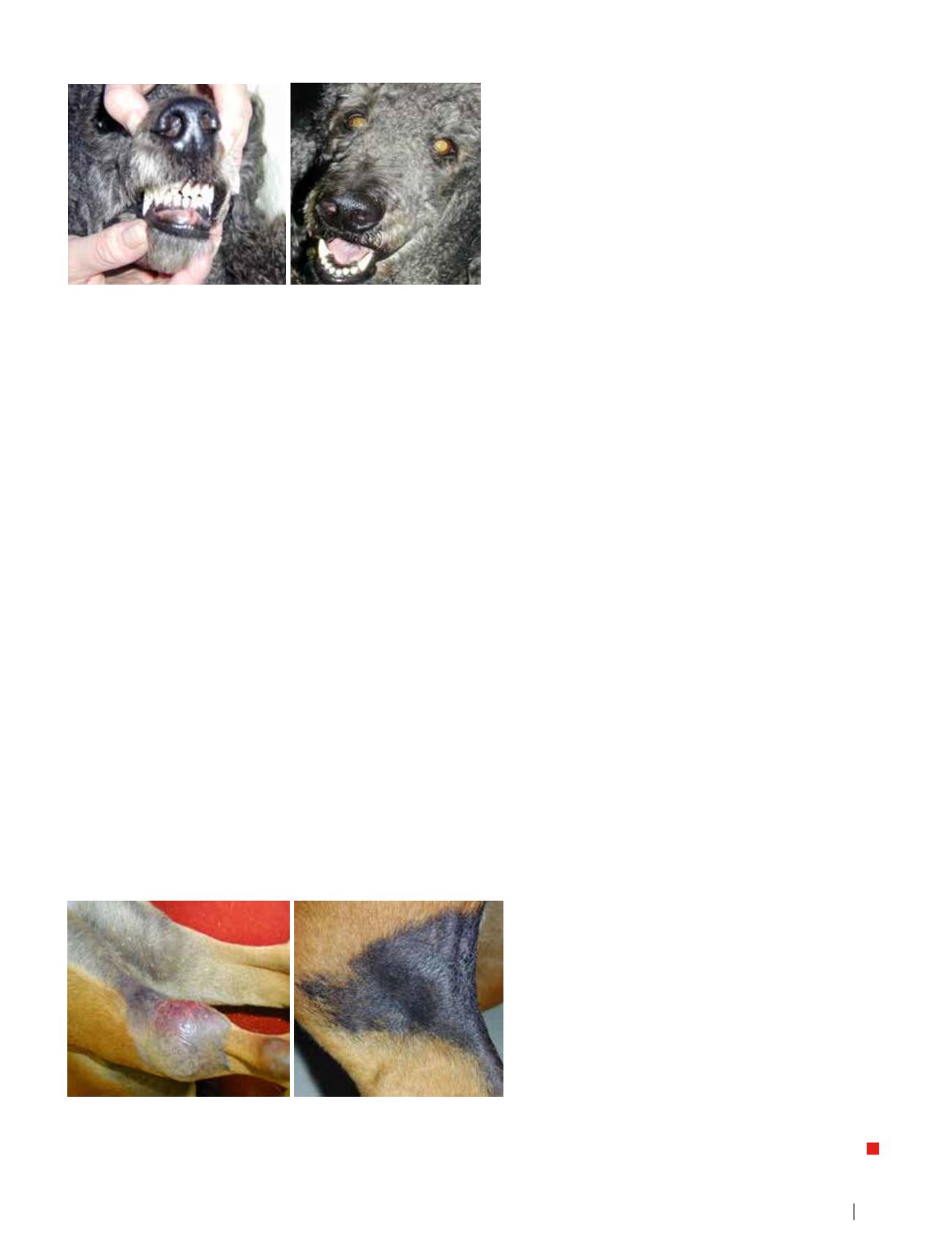
Mast Cell Tumour, Grade 3
The first photograph shows the MCT at presentation; the second
photograph was taken three years later, after radiation, surgery and post-
surgery radiation (total radiation 10 x 3 = 30 Gys)
Histiocytoma
The first photograph shows the tumour at presentation, patient not
eating. Treated with radiation therapy (12 x 2 Gys). Second photograph
taken 80 months later, no recurrence.
Lead Article
I Hoofartikel
C
ancer
T
reatment
<<< 6