


 16 / 258


1326
G
ill
et al
.:
J
ournal of
AOAC I
nternational
V
ol
.
99, N
o
.
5, 2016
where Result D is the vitamin D
2
or vitamin D
3
concentration
in the sample (μg/h), PA
NLD
is the peak area of vitamin D
2
or vitamin D
3
in the sample, PA
SILD
is the peak area of
d6
-
vitamin D
2
or
d6
-vitamin D
3
in the sample, SILISD
concn
is the
concentration of
d6
-vitamin D
2
or
d6
-vitamin D
2
in the SILIS
(ng/mL), L is the slope of the calibration curve, SILIS
alqt
is the
volume of the SILIS aliquot spiked into the sample (0.5 mL),
S
mass
is the mass of the sample (g), 1000 is the mass conversion
factor (ng/g to μg/g), and 100 is the mass conversion factor
(μg/g to μg/hg).
(n)
The concentration (w/v) of vitamin D
2
or vitamin D
3
in
ready-to-feed (RTF) liquids is calculated as
=
×
×
×
Result D
PA
PA
SILIS
L
SILIS
S
100
1000
NLD
SILD
Dconcn
alqt
vol
where Result D is the vitamin D
2
or vitamin D
3
concentration
in the sample (μg/dL), PA
NLD
is the peak area of vitamin D
2
or vitamin D
3
in the sample, PA
SILD
is the peak area of
d6
-
vitamin D
2
or
d6
-vitamin D
3
in the sample, SILIS
Dconcn
is the
concentration of
d6
-vitamin D
2
or
d6
-vitamin D
2
in the SILIS
(ng/mL), L is the slope of the calibration curve, SILIS
alqt
is the
volume of the SILIS aliquot spiked into the sample (0.5 mL),
S
vol
is the volume of the sample (g), 1000 is the mass conversion
factor (ng/g to μg/g), and 100 is the mass conversion factor
(μg/g to μg/hg).
(o)
The concentration of vitamin D
2
or vitamin D
3
as IU/hg
in the sample is calculated as
Result IU hg Result g/hg 40
(
)
(
)
=
µ ×
where 40 is the dietary conversion factor (μg/hg to IU/hg).
K. Data Handling
Report results as μg/hg to one decimal place or as IU/hg to
zero decimal places.
Results and Discussion
Method Optimization
The advantages of using the described derivatization strategy
for the analysis of vitamin D are that many compounds (such as
plant sterols) that are isobaric with vitamin D
2
and vitamin D
3
are excluded from detection because they lack the conjugated
diene structure, and therefore do not form adducts. The
derivatization of vitamin D with PTAD produces two epimers,
6S and 6R, because PTAD reacts with the
cis
-diene moiety from
both the
α
-side and the
β
-side, with the ratio of 6S:6R being
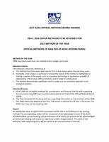
approximately 4:1 (6). The 6S/6R epimers coelute using the
described chromatographic conditions, and the typical MRM
chromatograms for a sample are shown in Figures 1 and 2.
Figure 1. MRM chromatogram for vitamin D
2
.
Figure 2. MRM chromatogram for vitamin D
3
.
16