
Chapter 2
•
Cardiovascular Care
21
vessel that leads to the heart. A
contrast dye visible in x-rays is
injected through the catheter
and flows through the heart
arteries to search for narrowed
or blocked coronary arteries
called a coronary angiography or
coronary arteriography. Cardiac
catheterization is performed to:
•
Identify diseases of the heart
muscle, valves, or coronary (heart)
arteries
•
Measure the pressure in the
chambers of the heart
•
Measure the oxygen content in the
chambers of the heart
•
Evaluate the ability of the
pumping chambers to contract
•
Look for defects in the valves or
chambers of the heart
•
Myocardial biopsy
Echocardiography
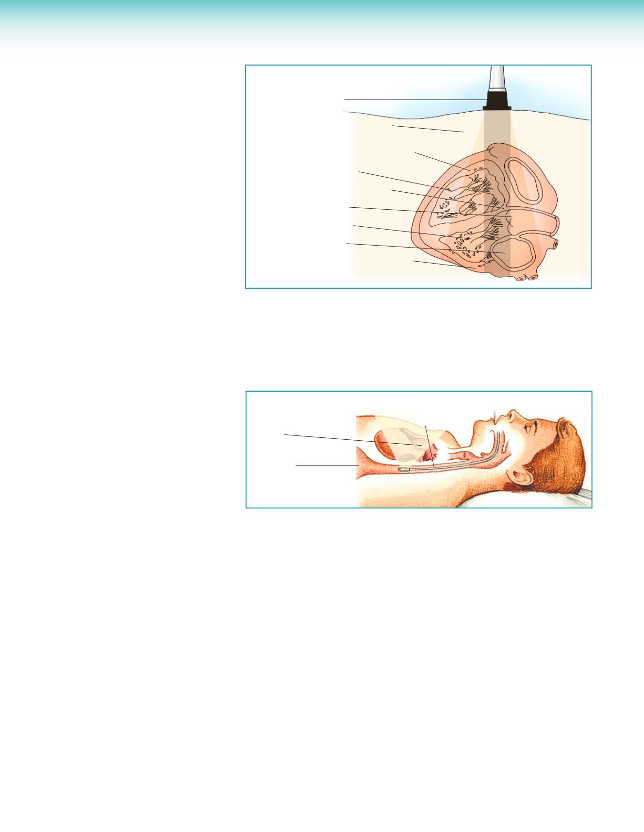
Echocardiography is an excellent
real-time imaging technique with
a high degree of clinical accuracy.
Echocardiograph uses ultra-high
frequency sound waves to help
examine the size, shape, and motion
of the heart’s structures. A special
transducer is placed over the patient’s
chest over an area where bone and
tissues are absent. It directs sound
waves to the heart structures and
converts them to electrical impulses.
These electrical impulses are sent
to the echocardiograph and
displayed on a screen. The image
is then recorded on a strip or
videotaped.
Transesophageal
Echocardiography
Transesophageal echocardiography
(TEE) produces pictures of the
heart using high-frequency sound
waves (ultrasound). Unlike a
standard echocardiogram, the
echo transducer that produces the
sound waves for TEE is attached to
a thin tube that passes through the
mouth and down the throat into the
esophagus. Because the esophagus
is so close to the upper chambers of
the heart, very clear images of those
heart structures and valves can be
obtained. TEE is used when more
Transducer
Anterolateral chest wall
Right ventricular anterior wall
Right ventricle
Intraventricular septum
Aortic valve
Left ventricle
Left atrium
Left ventricular posterior wall
detailed information is required than
a standard echocardiogram can give
them. The sound waves sent to the
heart by the probe in the esophagus
are translated into pictures on a
video screen. TEE visualizes the
heart’s structure and function and
provide clearer pictures of the upper
chambers of the heart, and the
valves between the upper and lower
chambers of the heart, than standard
echocardiograms.
Aorta
Stomach
Esophagus
Cardiac Magnetic
Resonance Imaging
Magnetic resonance imaging (MRI) is
a noninvasive test that uses a magnetic
field and radiofrequency waves to
create detailed pictures of organs and
structures. It can be used to examine
the heart and blood vessels, and to
identify areas of the brain affected
by stroke. MRI is also sometimes
called nuclear magnetic resonance
(NMR) imaging. MRI uses a powerful
magnetic field, radiofrequency waves,
and a computer to create detailed
cross-sectional (2-dimensional) and
3-dimensional images of the inside
of the body without using ionizing
radiation (like x-rays, computed
tomography, or nuclear imaging).
This test can identify the heart’s
structure (muscle, valves, and
chambers) and how well blood flows
through the heart and major vessels.
MRI can identify damage to the
heart from an MI, or if there is lack
of blood flow to the heart muscle
because of narrowed or blocked
arteries.
MRI is useful in identifying:
•
Tissue damage
•
Reduced blood flow in the heart
muscle
•
Aneurysms
•
Diseases of the pericardium
•
Heart muscle diseases, such as
heart failure (HF) or enlargement
of the heart, and tumors
•
Heart valve disorders
•
Congenital heart disorders
•
Success of surgical repair


