


 4 / 37


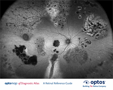
af
Diagnostic Atlas
A Retinal Reference Guide
Autofluorescence
2
1. Holz, F. S.-V. (2010). Atlas of Fundus Autofluorescence Imaging. Heidelberg, Germany: Springer-Verlag.
2. Delori, F. G. (2001). Age-Related Accumulation and Spatial Distribution of Lipofuscin in RPE of Normal Subjects. IVOS, 42(8), 1855-1866.
3. Sadda, S. (October 2013). Evaluating Age-Related Macular Degeneration With Ultra-widefield Fundus autofluorescence. Retina Today.
opto
map
af
(autofluorescence) is a non-invasive, in-vivo imaging modality used to provide
information on the health and function of the retinal pigment epithelium (RPE).
Over time, the retinal photoreceptors naturally age and produce a metabolic waste known
as lipofuscin. Lipofuscin is the fatty substance found in the retinal pigment epithelium.
Excessive amounts can be caused by the aging retina, certain retinal diseases and/or the
progression of diseases.
1
It has been thought that excessive levels of lipofuscin could aect
essential RPE functions that contribute to the progression of age-related macular degeneration
(AMD).
2
These findings have also been shown to have prognostic value and help to predict
which eyes are at greater risk of progression to advanced disease.
3
Typically, autofluorescence imaging has clinical applications in age-related macular degeneration,
central serous retinopathy, choroidal tumors and nevi, inflammatory diseases, inherited
disease, optic nerve head drusen, pattern dystrophies, retinal toxicity and retinal detachments.
Autofluorescence excitation wavelength is between 480-510 nm, with an emission wavelength
from 480-800 nm.
1
opto
map
af
uses a wavelength of 532nm to capture an image.