


 8 / 37

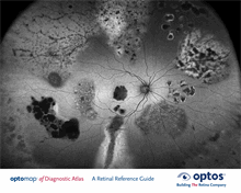
Vogt-Koyanagi-Harada
Macular Dystrophy
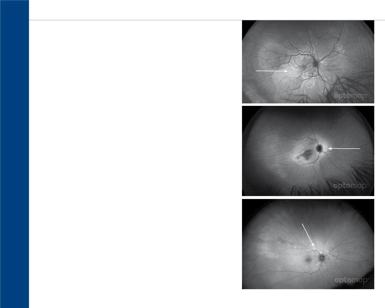
Angioid Streaks
Hyperautofluorescence
6
Hyperautofluorescence
is an increased AF signal which will appear white on the
image. Many disease states can cause the accumulation
of lipofuscin and a hyperautofluorescence signal
1
:
∙Stargardts disease
∙Best disease
∙Adult vitelliform macular dystrophy
∙Age-related macular degeneration
∙Intraretinal fluid (e.g., macular edema)
∙Subretinal fluid
∙Choroidal tumors and melanomas
∙Drusen
∙Older Intraretinal and subretinal hemorrhages
∙Choroidal vessels in the presence of RPE and
choriocapillaris atrophy (e.g., the center of laser
scars or within patches of RPE atrophy)
∙Idiopathic macular telangiectasia
∙Cystoid macular edema
∙Optic Nerve Head Drusen
1. Schmitz-Valckenberg, S. H. (2008). Fundus Autofluorescence Imaging. Retina, The Journal of Retinal and Vitreous Diseases, 28(3), 385-409.