


 6 / 37


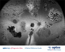
af
Diagnostic Atlas
A Retinal Reference Guide
Vein
is any of the tubes forming part of the
blood circulation system of the body,
carrying in most cases oxygen-depleted
blood toward the heart.
Macula
is a small central area of the
retina surrounding the fovea;
area of acute central vision.
Fovea
is the central pit in the macula that
produces sharpest vision. It contains
a high concentration of cones and no
retinal blood vessels.
Optic Disc,
Optic Nerve Head (ONH)
is the ocular end of the optic nerve.
It denotes the exit of retinal nerve
fibers from the eye and entrance
of blood vessels to the eye.
Artery
is any of the muscular-walled tubes
forming part of the circulation
system by which blood (mainly that
which has been oxygenated) is
conveyed from the heart to all parts
of the body.
Retinal Nerve Fiber Layer
(RNFL)
is the expansion of the fibers of
the optic nerve; it is thickest near
the nerve diminishing toward the
ora serrata.
The Retina
is the light-sensitive layer of tissue that lines the inside of the eye
and sends visual messages through the optic nerve to the brain.
The Choroid
is the vascular (major blood vessel) layer of the eye
lying between the retina and the sclera. It provides
nourishment to outer layers of the retina to the brain.
Retinal Anatomy
4