


 10 / 44

and then turned off. Iterating this process hundreds of times
builds up a pointillist image of the sample, with resolution
far below the diffraction limit
( 8). This advance was recog-
nized in the 2014 Nobel Prize in Chemistry.
Highlighting
Photoactivatable and photoswitchable FPs have served as
optical highlighters for tracking the flow of matter in a
cell. One can tag a cellular structure with a flash of light
and then follow the motion of that structure through the
cell. This enables one to probe how mitochondria move
through neurons and track the assembly and disassembly
of microtubules.
Binding
Many nonfluorescent proteins change shape when they bind
a ligand or a partner. The chromophore in most FPs must
pack snugly among surrounding amino acids to fluoresce.
Crack open the protein barrel or expose the chromophore
to water, and the fluorescence goes away. This combination
of features has been exploited by constructing circularly
permuted FPs in which the two ends of the amino acid chain
are linked, and a new break is introduced near the chromo-
phore. A slight tug on the new ends of the chain can revers-
ibly disrupt the fluorescence and, by fusing nonfluorescent
sensor domains to circularly permuted FPs, one can make
fluorescent sensors that report ATP, calcium, membrane
voltage, and ligand binding to G protein-coupled receptors.
The most dramatic applications of fluorescent sensor pro-
teins come from the GCaMP family of Ca
2
þ
indicators
( Fig. 2B
). The concentration of this ion blips upward every
time a neuron fires. Expression of GCaMP-based reporters
in the brains of worms, flies, fish, and mice has led to spec-
tacular movies of the coordinated activation patterns of
thousands of neurons.
Within the last year, scientists have started to engineer
more complex combinations of functions into GFP-based
optogenetic tools. For instance, the calcium-modulated pho-
toactivatable ratiometric integrator (CaMPARI) protein
starts life as a fluorescent calcium indicator, and, in the
simultaneous presence of neural activity and violet illumi-
nation, converts from green to red
( 9). This behavior lets
one record a photochemical imprint of the calcium level
in a large volume of tissue at a defined moment in time.
One can then image the tissue at leisure, with high resolu-
tion in space, to map this snapshot of activity.
Many new types of sensors are still needed. A fluorescent
reporter for glutamate has been described
( 10), but reporters
for many other neurotransmitters (gamma-aminobutyric
acid, dopamine, serotonin, and acetylcholine) are still in
development. It also is challenging to sense physical forces.
Fluorescent reporters for membrane voltage
( 11) and cyto-
skeletal tension
( 12) have been developed, but we lack
voltage indicators that perform well enough to be used
in vivo or that can be targeted to intracellular membranes
(mitochondria, vesicles, and endoplasmic reticulum). We
also lack fluorescent reporters for many of the subtle, but
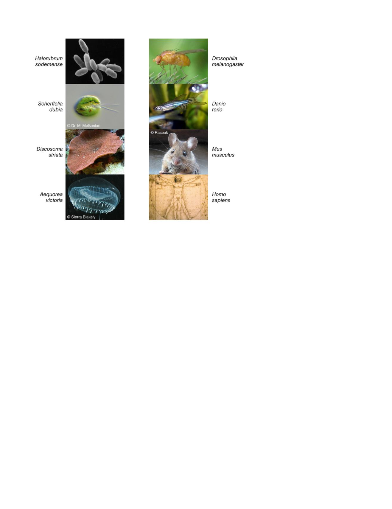
FIGURE 1 Optogenetic mix-and-match. (
Left
)
Organisms whose genes have yielded new optoge-
netic tools. (
Right
) Organisms into which scientists
have transferred these genes. To see this figure in
color, go online.
Biophysical Journal 110(5) 997–1003
998
Cohen