


 11 / 44

likely important, physical forces in biology: plasma mem-
brane tension, osmotic pressure, or stresses between cells
and their neighbors or the surrounding extracellular matrix.
In the applications described above, light interacts with
the FPs—eliciting fluorescence, changing the brightness,
or changing the color—but the light does not fundamentally
change the underlying biological process (at least not inten-
tionally; phototoxicity is a constant concern for these exper-
iments). The true power of optogenetics emerged when
scientists started to use light to perturb the underlying
biology in a precise way.
Microbial rhodopsins bring light to the membrane
Most living things sense and respond to changes in light.
A diverse set of transducers has evolved to couple light
into biochemical signals. Here, we focus on the microbial
rhodopsins as a paradigmatic example. The first microbial
rhodopsin was discovered in the early 1970s in a halophilic
archaeon,
Halobacterium salinarum
, in the salt marshes of
San Francisco Bay. The protein has seven transmembrane
a
helices and a retinal chromophore covalently bound in
its core. Upon illumination, the retinal undergoes a trans-
to-
cis
isomerization, which induces a series of shape
changes in the protein that lead to pumping of a proton
from inside the cell to the outside. The protons return
back into the cell through the ATP synthase, powering the
metabolism of the host.
More than 5000 types of microbial rhodopsins have
been identified by metagenomic sequencing. They are
found in archaea, prokaryotes, and eukaryotes. Most are
uncharacterized, and these proteins mediate a huge variety
of interactions between sunlight and biochemistry. Some
act as light-driven proton pumps (e.g., bacteriorhodopsin,
proteorhodopsins, and archaerhodopsins) and others act
as light-driven chloride pumps (e.g., halorhodopsin),
light-activated signaling molecules (sensory rhodopsins),
or light-gated cation channels (channelrhodopsins).
The discovery that channelrhodopsin 2, derived from the
green alga
Chlamydomonas reinhardtii
, functioned as a
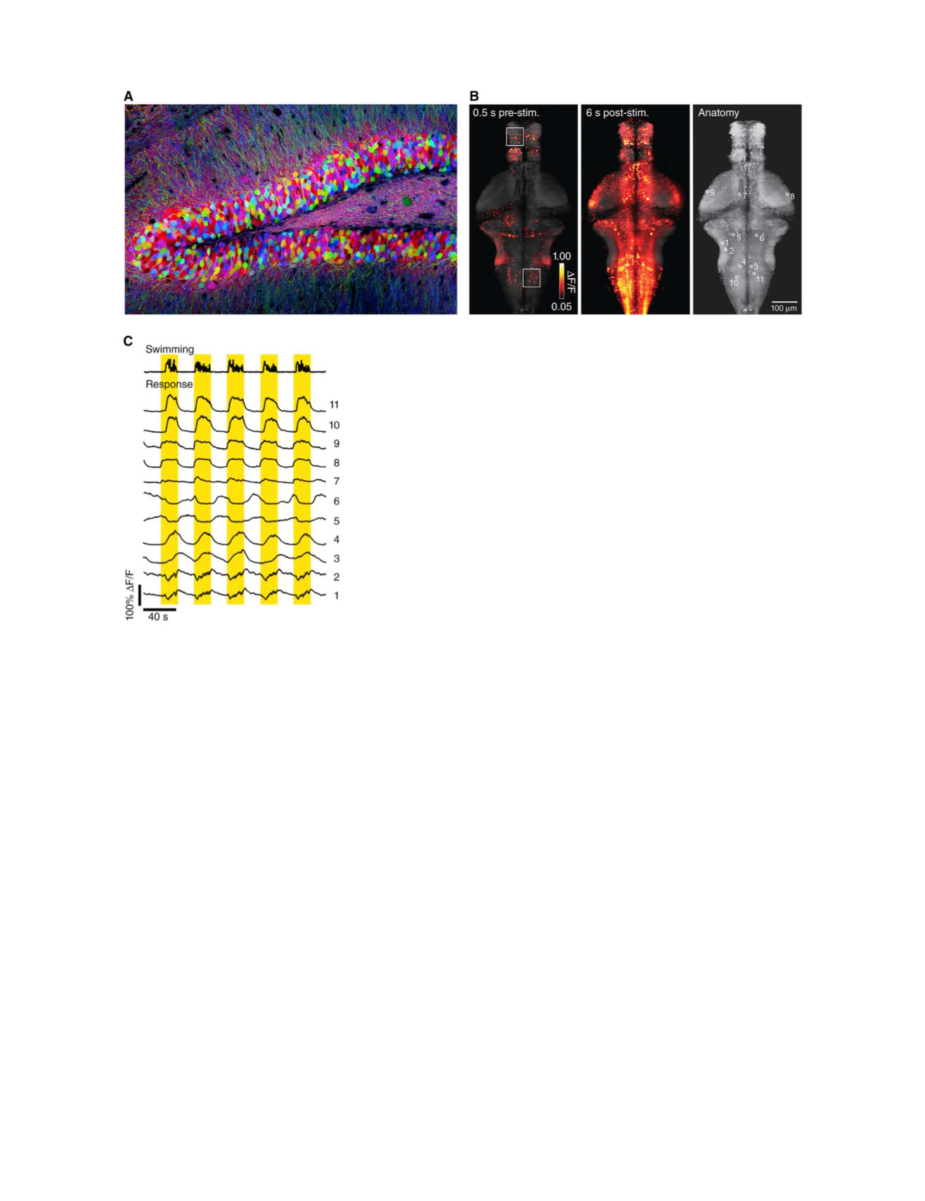
FIGURE 2 Fluorescent protein-based sensors light up the brain. (
A
) Brainbow mouse hippocampus. Random genetic recombination events turn on a
different subset of fluorescent proteins in each neuron, giving each one a unique hue. (
B
) Imaging neural activity in the brain of a zebrafish via the calcium
indicator GCaMP6s. The fish was immobilized over an image of a drifting grating. When the grating started to move (
stim
), the fish tried to swim to maintain
its position within the visual field. The central image shows the brain regions that activated when the fish swam. (
C
) Activity of neurons indicated in the right
panel of (
B
) during swimming. (
A
is from Jeff Lichtman;
B
and
C
are from
( 23).) To see this figure in color, go online.
Biophysical Journal 110(5) 997–1003
Optogenetics
999