


 12 / 44

light-gated cation channel triggered a race to apply this pro-
tein to control neural firing with light. A first-person histor-
ical account has been written by one of the chief
protagonists, Ed Boyden
( 13), and a thorough review of
the early literature has also been written by Karl Deisseroth,
another key protagonist, and his colleagues
( 14). Early dem-
onstrations in cultured neurons were quickly followed by
demonstrations in mouse brain slice, chick spinal cord,
worms, flies, and zebrafish. Control of rodent behavior
started with simple whisker movements, but then quickly
expanded to control of locomotion, sleep, feeding, aggres-
sion, memory, and social interactions
( Fig. 3A
).
Recent work on pup rearing demonstrates the sophisticat-
ion and precision that optogenetic stimulation has reached
( 15). In male or female mice showing parenting behavior,
a subpopulation of neurons became active in the medial pre-
optic area. These cells were genetically targeted with a Cre-
dependent channelrhodopsin construct and, in virgin male
mice that normally show aggression toward pups, optoge-
netic actuation reversibly switched the animals into a
grooming mode. These and many other optogenetic experi-
ments demonstrate that seemingly complex rodent behav-
iors can be elicited by precise actuation of relatively small
numbers of neurons in genetically defined circuits.
Converting microbial rhodopsins into reporters
Efforts to engineer better optogenetic neural modulators
relied heavily on mechanistic insights obtained from de-
cades of detailed biophysical studies of the photocycle of
bacteriorhodopsin and its homologs. A standard technique
in this arena was transient absorption spectroscopy, which
triggers the photocycle with a flash of light and subsequently
records absorption spectra as a function of time. Motion of
the proton through the protein core was accompanied by
shifts in the absorption spectrum.
My lab discovered that microbial rhodopsin proton
pumps are weakly fluorescent and that this fluorescence
varies depending on the location of a proton in the core of
the protein. Changes in membrane voltage could reposition
the proton, thus changing the fluorescence. We realized that
this phenomenon might provide a novel route toward one of
the longest-standing challenges in neuroscience: to develop
a fast and sensitive optical reporter of membrane voltage.
The initial proteorhodopsin-based voltage indicator
(called PROPS) functioned only in bacteria and led to the dis-
covery that
Escherichia coli
generate spontaneous electrical
spikes
( 16). Neither the underlying mechanism nor the bio-
logical function of this spiking is well understood. Of the
millions of species of bacteria in the world, we know almost
nothing about the electrophysiology of any of them.
PROPS did not work in mammalian cells because the pro-
tein did not traffic to the plasma membrane. After an unsuc-
cessful year-long effort to engineer membrane trafficking
into PROPS, we switched to Archaerhodopsin 3 (Arch), a
protein derived from a Dead Sea microorganism,
Haloru-
brum sodomense
, which was discovered in the early 1980s
by an Israeli microbiologist. Arch immediately showed
voltage-sensitive fluorescence in mammalian cells
( 17).
Further protein engineering eliminated the photocurrent
and improved the sensitivity and speed of the protein, lead-
ing to the QuasAr family of voltage indicators (
18 ).
By good fortune, the fluorescence of microbial rhodop-
sins is excited by red light and emits in the near infrared.
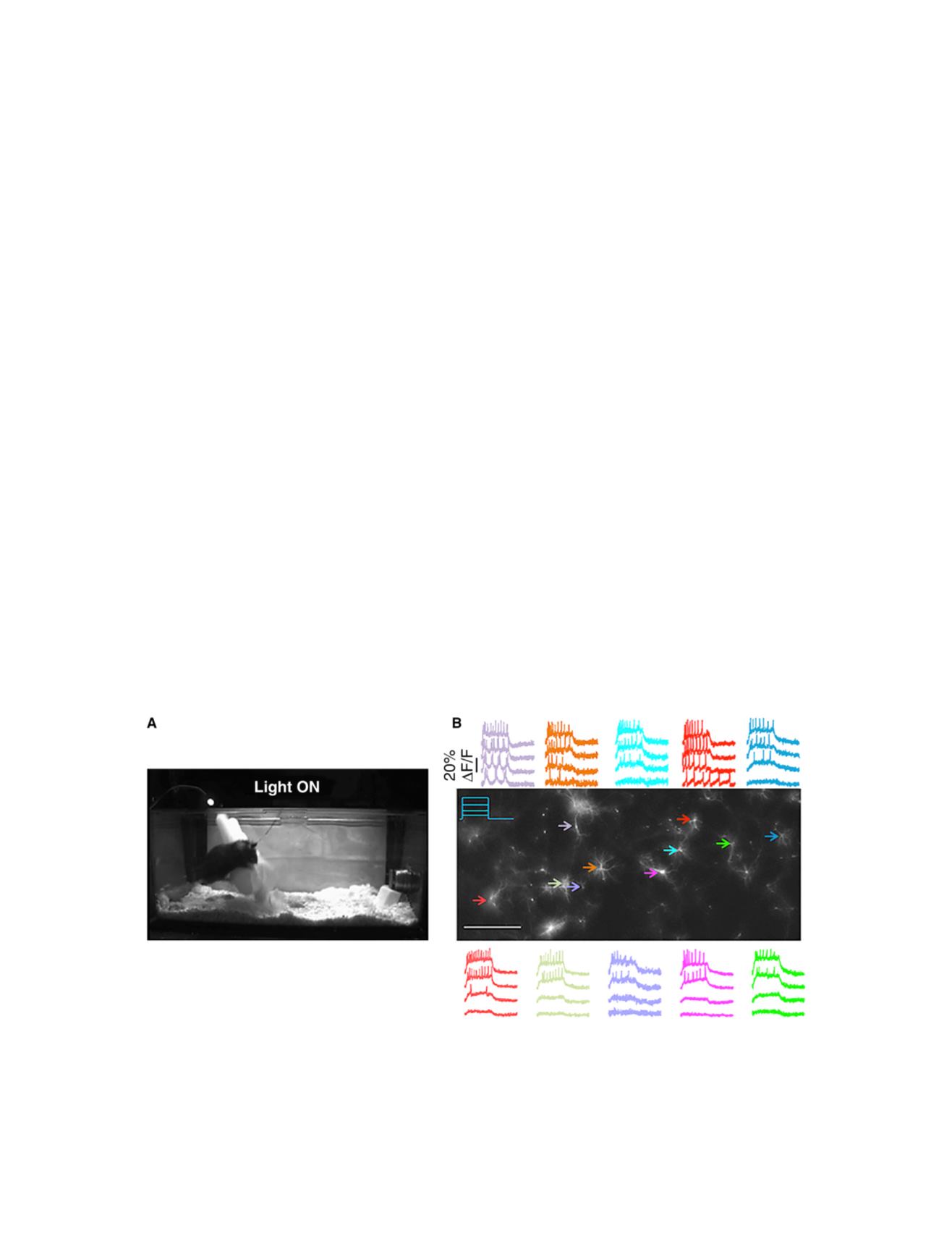
FIGURE 3 Optogenetic control and readout of neural activity. (
A
) Optogenetic control of aggression. In this still from a movie, the mouse is expressing
Channelrhodopsin 2 in its ventromedial hypothalamus, ventrolateral subdivision (VMHv1). Illumination of this region through an optical fiber causes the
animal to attack an inflated rubber glove, which it would otherwise ignore. (
B
) All-optical electrophysiology. These rat hippocampal neurons express the
Optopatch constructs, comprising a blue light-activated channelrhodopsin variant (CheRiff) and a red light-activated voltage indicator (QuasAr2). Illumina-
tion with 500 ms pulses of blue light triggers intensity dependent neural activity. Scale bar, 0.5 mm. (
A
is from
( 24);
B
is from
( 25).) To see this figure in color,
go online.
Biophysical Journal 110(5) 997–1003
1000
Cohen