


 3022 / 3581

IGRT for Head and Neck
Coen Rasch
Head and Neck
Delineation of GTV in H&N
10 patients with NPC (cT2b – cT4, Nx)
10 Observers from 6 institutes in NL, D
and US
Phase I
– Delineation of GTV on CT
n of GTV on CT
– Diagnostic MRI copy available
Phase II, after > 1 year
– Improved delineation protocol
– Delineation on co-registered CT/MRI
– Computer aided delineation tool (Snake
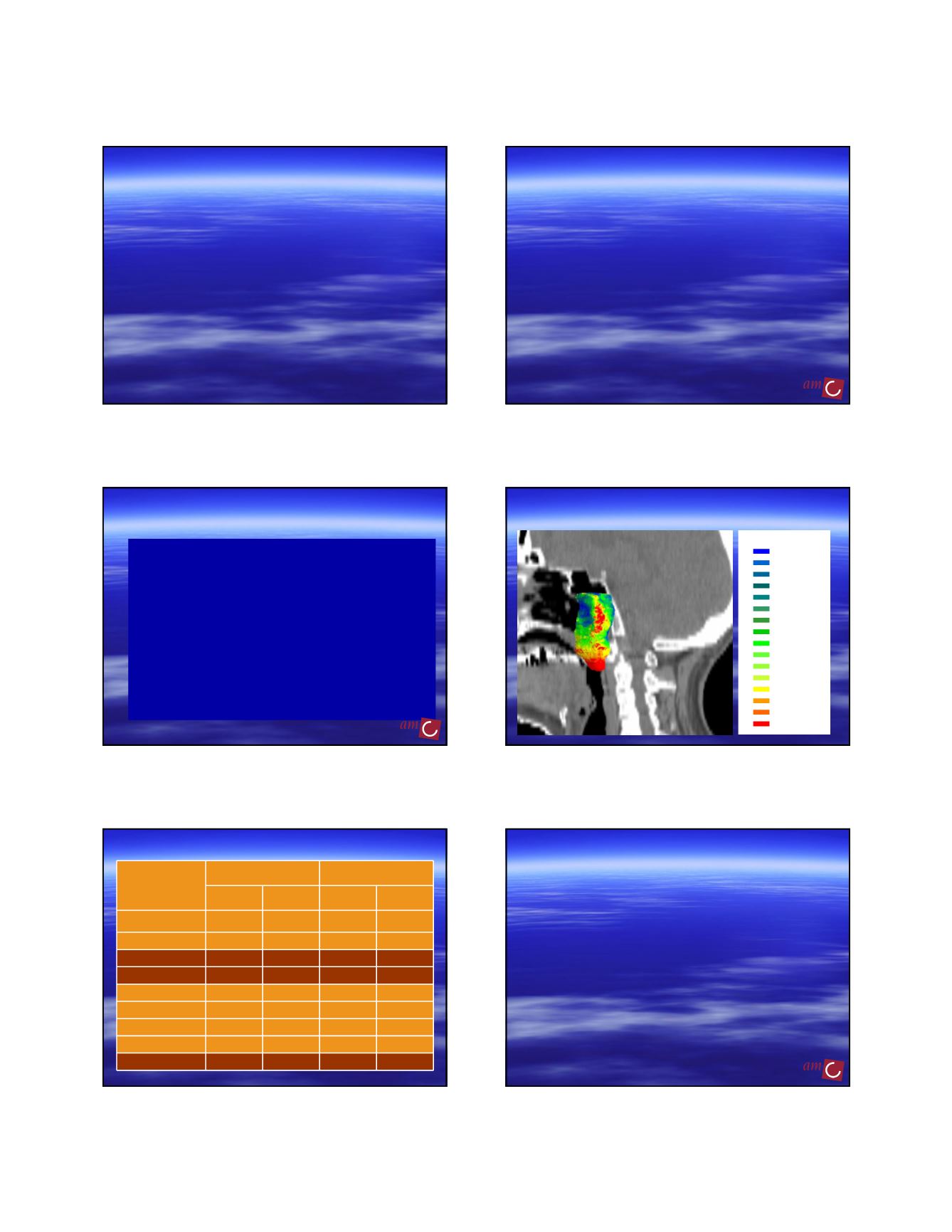
3-D median surface with local SD
0.0 – 0.5
0.5 – 1.0
1.0 – 1.5
1.5 – 2.0
2.0 – 2.5
2.5 – 3.0
3.0 – 3.5
3.5 – 4.0
4.0 – 4.5
4.5 – 5.0
5.0 – 5.5
5.5 – 6.0
6.0 – 6.5
6.5 – 7.0
7.0 – 7.5
> 7.5
LOCAL SD (mm)
Overall observer variation (SD)
Anatomical regions
Phase 1
Phase 2
SD CT (mm)
Agreement
(%)
SD CT/MRI
(mm)
Agreement
(%)
All regions 4.4 36 3.3 64
Anterior – Air
3.4
62
2.7
79
Dorsal – Bone
3.6
49
2.7
84
Contra lateral
4.2
16
3.5
66
Pterygoid M.
4.3
35
3.1
61
Parapharyngeal
4.4
31
3.3
59
Soft Palate
4.7
37
3.0
67
Sphenoid
5.0
28
4.2
48
Caudal side
7.7
5
3.3
56
Delineation effect on dose in
conformal and IMRT plans
Paranasal sinus cancer, nine patients
Two observers
– Elective CTV (described in anatomical terms)
Mean ratio 0.9
– Boost CTV (the tumor plus margin)
Mean ratio 2.6
Two treatment planners
– IMRT
– 3D conformal
•Rasch et al IJROBP 2002