


 231 / 258


F
eng
et al
.:
J
ournal of
AOAC I
nternational
V
ol
.
100, N
o
.
2, 2017
513
and BSA peak; perform a manual integration from the valley
between the end of the κ-casein peak and the peak of Ig H and
BSA to the end of the last peak in the e-grams (at least 9 min
after the peak of the 10 kDa IS).
(j)
To determine the casein region, set the start time for
casein integration just before the β-casein peak in the e-gram
of the SMP (about 3.1 min after the peak of the 10 kDa IS).
Referencing the SMP, identify the β-casein peak in the infant
formula samples, then set the start time of the casein region in
the infant formula to just before the β-casein peak. Set the end
time at the valley between the end of the κ-casein peak and
the Ig H and BSA peak (about 7.0 min after the 10 kDa IS).
F. Calculations
(a)
To calculate whey protein content, separately sum
the peaks in the following three regions: two at each end of
the e-gram (smaller and larger whey proteins) and one in the
middle. The middle region corresponds to casein proteins (A
cn
),
and the two others are summed together to obtain the whey
proteins (A
w
).
(b)
Whey protein content is calculated using the following
equations:
Percentage of whey protein
A
A A
w,c
w,c
cn
=
+
(1)
A A 1.4
w,c
w
= ×
(2)
where A
w
= total integrated areas of whey components;
A
w,c
= corrected integrated area of whey components; A
cn
= integrated area of casein components; and 1.4 = CF to account
for the difference between the mass-to-area ratio of whey
and casein proteins.
Results and Discussion
Specificity
(a)
Reagent blank
.—Each sample sequence was started
with purified water as a blank. The blank e-gram is shown
in Figure 1 (gray line). No peaks were detected after the
10 kDa peptide internal standard (IS). There was no significant
interference from other components in the protein region.
(b)
Placebo test
.—To test for the presence of interference
from nonprotein components in infant formulas, a placebo
infant formula trial sample that contained all of the ingredients
that are typical first-age formulas, except protein (vitamins,
minerals, fat, and carbohydrates), was manufactured. SDS-CGE
did not detect significant peaks at any of the protein regions in
the e-gram (Figure 1, black line).
(c)
Specific protein migration time and migration pattern of
whey proteins and caseins
.—The SDS-CGE method can separate
individual whey and casein protein standards very well, as
demonstrated with standard solutions containing five major whey
proteins (Figures 2 and 3) or four casein proteins (Figure 4), as
well as with fresh raw milk (Figure 5). Protein phosphorylation
and glycosylation delay casein migration times relative to their
molecular sizes (Figure 6). Protein glycation—the nonenzymatic
sugar modification of amines and the early stage of a Maillard
reaction—occurs during the mixing and heating of milk proteins
with lactose (3), which results in the splitting of several individual
milk proteins into several peaks representing the modified protein
glycoforms. This was seen for α-Lac and β-Lg, where splitting
was observed in a commercial sweet whey protein ingredient
(Figure 7) and by comparing the casein peaks in fresh milk and
in an SMP sample (Figures 5 and 8). Although glycation prevents
the complete separation of all proteins individually, whey proteins
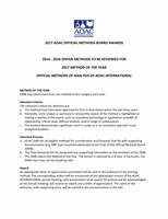
Figure 2016.15B. Separation of the protein MW size standard.
231