


 231 / 1082

S218
ESTRO 36 2017
_______________________________________________________________________________________________
M.T. G arcia-Hernandez
1
, B. Sanchez-Nieto
2
, J. Roselló
Ferrando
1
, L. Irazola
3
, J.A. Terrón
3
, F. Sanchez-Doblado
3,4
1
Hospital General Universitario de Vale ncia. ERESA,
Servicio de Radiofísica., Valencia, Spain
2
Instituto Física Pontificia Universidad Católica de Chile,
Departamento Física Médica, Santiago de Chile, Chile
3
Universidad de Sevilla, Departamento de Fisiología
Médica y Biofísica, Sevilla, Spain
4
Hospital Universitario Virgen Macarena, Servicio de
Radiofísica, Sevilla, Spain
Purpose or Objective
The study of Secondary Malignant Neopl asms, as a
consequence of the peripheral doses received by photon
radiotherapy patients, is becoming a topic of growing
interest due to the higher healing rates and life
expectancy accomplished nowadays. Two models have
been developed to estimate peripheral doses due to
photon and neutron dose deposition (1,2). The aim of this
work is the validation of a generic peripheral photon dose
model (1) for the flattening filter free (FFF) modality,
which was not originally considered.
Material and Methods
Measurements were carried out in a Varian Truebeam linac
for FF and FFF beams (with 6 and 10 MV) for two different
field sizes (3x3 and 10x10 cm
2
) with both, single (gantry
0º) and multiple incidences (0°, 45°, 90°, 135°, 180°,
225°, 270°, 315º). A CC13 (Iba Dosimetry) ionization
chamber was placed at a range of out-of-field distances
(10 to 60 cm from the field-edge) in a water-equivalent
phantom and irradiated with 1000 MU. The obtained
results for FF and FFF were compared to estimations with
the original model (1).
Results
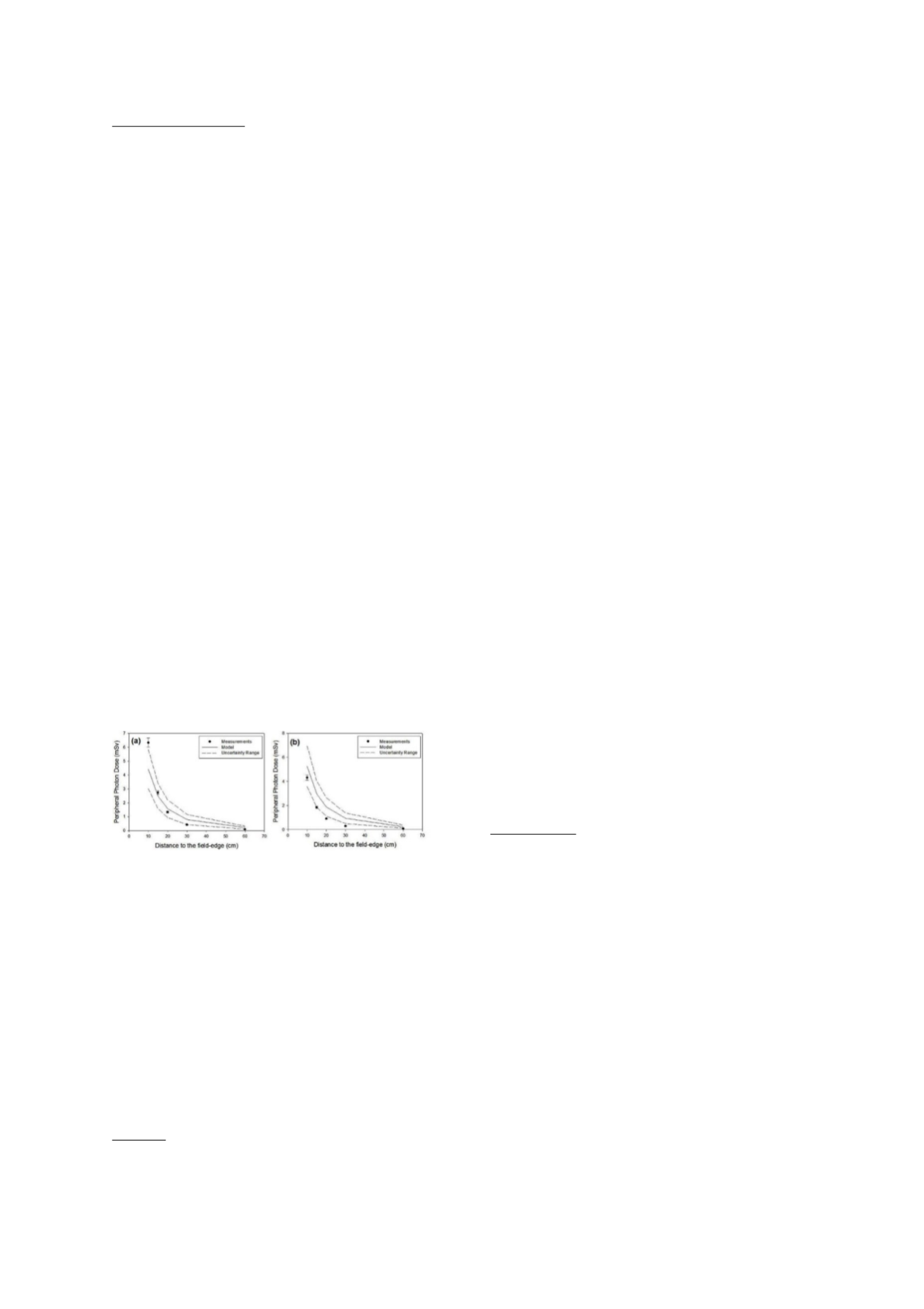
Experimental measurements, together with model
predictions for all combinations described were collected.
By way of example, results the 10x10 cm
2
field using 6 and
10 MV FFF multiple incidences have been depicted in Fig
1a and 1b, respectively. The uncertainty range (UR) of the
model (95% confidence interval) (1) as well as the 5%
uncertainty
estimated
for
the
experimental
measurements, are shown.
Conclusion
The original photon model tends to overestimate
peripheral doses, especially for the high energy. This is
due to the fact that FFF beams, in comparison with FF
beams, are associated to lesser scatter in the linac hea.
This effect is enhanced at higher energies. Thus, despite
that the original model could be used for peripheral
photon dose assessment in FFF modality (experimental
data are almost included within the UR of the model),
further investigation should be conducted to better model
the effect of the absence of the flattening filter.
References
(1) Sánchez-Nieto et al
.,
Biomed Phys Eng Express
2015;1:045205.
(2) Sánchez-Doblado et al.,Phys Med Biol 2012;57:6167–
6191.
PV-0418 Characterisation of the prototype plastic
scintillation detector (PSD) in a strong magnetic field
M. Asghar
1,2,3
, W. Zhifei
1
, Y. Suh
1
, D. O'Brien
1
, S. Beddar
1
,
S.A. Buzdar
3
, G.S. Ibbott
1
1
The University of Texas MD Anderson Cancer Center,
Department of Radiation Physics, Houston, USA
2
Bahawalpur Institute of Nuclear Medicine and Oncology
BINO, Department of Medical Physics, BAHAWALPUR,
Pakistan
3
The Islamia University of Bahawalpur, Medical Physics
Research Group- Department of Physics, BAHAWALPUR,
Pakistan
Purpose or Objective
Novel treatment units are becoming available that
combine a magnetic resonance (MR) imager with a mega-
voltage radiation beam. The magnetic field of the MR
Imaging affects the trajectories of secondary electrons
and influences the performance of several types of
radiation detectors, including ion chambers and diodes. A
dosimeter that is not influenced by the magnetic field
would be valuable for commissioning and quality
assurance (QA) of an MR-guided treatment unit. The
purpose of this work was to characterise the performance
of a PSD in the magnetic field of an MR-LINAC system.
Material and Methods
The MR-LINAC system consists of a 1.5 T Achieva MRI
system (Philips, Netherlands) and a 7 MV linear
accelerator (Elekta, UK). The feasibility of using the
prototype PSD (Standard Imaging, USA) in MR-LINAC
radiation therapy system was evaluated by investigating
possible effects of the strong magnetic field on the
performance characteristics of the PSD. The effects of
orientation, axial rotation symmetry, and optical
connectivity of the PSD and that of the photo-diode
position were measured in the presence and absence of
the magnetic field.
Results
The mean percent differences in the PSD signals for
different orientation of the PSD and for various axial
rotations of the PSD in the transverse magnetic field
between magnet ramped up and down were 1.33%
(±0.92%) and 1.37% (±1.01%), respectively. The effects of
optical connectivity and photo-diode position were
insignificant on the signal.
Conclusion
We conclude that the PSD can be used for dosimetry of the
MR-LINAC radiation therapy system as the effect of a
strong magnet field was insignificant on the
characteristics of the PSD investigated. It would be a good
detector for commissioning and QA of an MR-guided
system.
PV-0419 The impact that geometric variability in
ionization chamber construction has on kQQo
J. Puxeu Vaqué
1,2,3
, M. Duch Guillen
4
, M.C. Lizuain
Arroyo
3
, W.H. Nailon
2
1
Hospital Universitari Sant Joan de Reus, Servei de
Protecció Radiològica i Física Mèdica, Reus, Spain
2
Edinburgh Cancer Centre, Department of Oncology
Physics, Edinburgh, United Kingdom
3
Institut Català D'Oncologia, Servei de Física Mèdica i
Protecció Radiològica, L'hospitalet de Llobregat, Spain
4
Universitat politècnica de Catalunya, Institut de
Tècniques Energètiques, Barcelona, Spain
Purpose or Objective
To investigate the influence that geometric uncertainties
in the manufacturing process of three different ionization
chambers has on the beam quality correction factor
k
QQo.
Ionization chambers (IC) have been used as reference
detectors in clinical practice for decades. In 2000 a new
code of practice (TRS-398) was introduced based on the
calibration of the ionization chambers in terms of
absorbed dose to water instead of the previous code based
on air kerma determination (TRS-277).
Not all standard laboratories have beams with the same
user beam qualities. One common approach is for the
Standard Dosimetry Laboratory (SDL) to perform a
calibration of the user’s ionization chamber in the beam
quality of the Co-60 source. They may also provide