


 233 / 1082

S220
ESTRO 36 2017
_______________________________________________________________________________________________
Conclusion
The proposed method of analysis of isolated errors gives
the magnitude of errors. In general, the most important
are leaf gap width errors. Exception and further analysis
is required for VMAT patients with low DR. ArcCheck
positioning errors should not affect the gamma results
while being in specification tolerance.
PV-0421 In-magnet measurement setup for proof-of-
concept and commissioning of MR integrated proton
therapy
A. Lühr
1,2,3
, S. Gantz
1,3
, S. Schellhammer
1,3
, O. Zarini
4
, K.
Zeil
4
, U. Schramm
4
, A. Hoffmann
1,3,5
1
Helmholtz-Zentrum Dresden - Rossendorf, Institute of
Radiooncology, Dresden, Germany
2
German Cancer Consortium DKTK, Partner Site Dresden,
Dresden, Germany
3
OncoRay – National Center for Radiation Research in
Oncology, Medical Radiation Physics, Dresden, Germany
4
Helmholtz-Zentrum Dresden - Rossendorf, Institute of
Radiation Physics, Dresden, Germany
5
Faculty of Medicine and University Hospital Carl Gustav
Carus at the Technische Universität Dresden,
Department of Radiation Oncology, Dresden, Germany
Purpose or Objective
There is growing interest to explore the concept of
magnetic resonance integrated proton therapy (MRiPT).
However, no experimental proof-of-principle has been
established so far. The aim of this work was to develop an
in-magnet measurement setup that facilitates to
investigate the dosimetric feasibility of MRiPT and to
develop a commissioning procedure for future MRiPT
devices.
Material and Methods
A C-shaped 0.95 T permanent neodymium (NdFeB) dipole
magnet was used. The magnetic main and fringe fields
were characterized using 3D automated Hall probe
measurements. A method and procedure were established
to perform periodic quality assurance (QA) measurements
of the magnetic field’s constancy. A 3D vector field
representation for the magnetic flux density distribution
was calculated by finite-element modeling (FEM) using
COMSOL-Multiphysics®. For irradiation experiments,
proton beams of 80–225 MeV were collimated using brass
apertures having circular voids of either 5 or 10 mm
diameter. The beams entered a PMMA slab phantom being
placed inside the magnet’s horizontal air gap,
perpendicularly to the main field component. Proton
beam trajectories and depth-dose curves in the presence
of the magnetic field were measured with Gafchromic
EBT3 film, being placed between the two slabs of the
phantom. Reference trajectories were measured without
magnetic field. In transmission experiments without
phantom, beam deflections were measured with a 2D
scintillation detector (Lynx, IBA Dosimetry) positioned
perpendicular to the beam at 24 cm distally from the
magnet.
Results
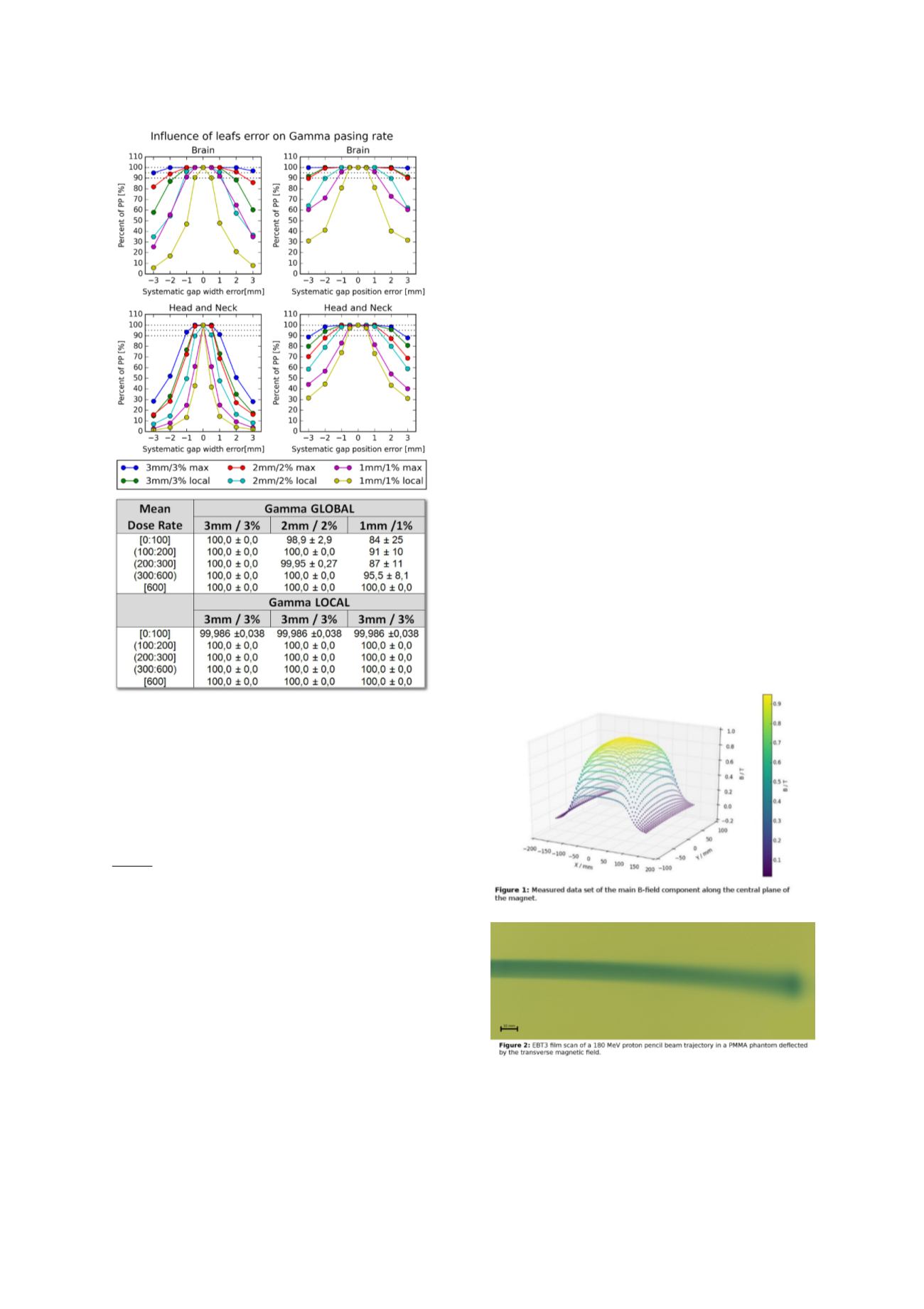
Magnetometry results (Fig. 1) validated the 3D magnetic
flux density distribution as calculated by FEM. The
simulation tended to underestimate the measured
magnetic field strength in the plateau area by about 2%
(mean difference 20 mT). In repeated QA measurements,
field strength changes remained below a threshold of 3
mT. For all proton energies, the lateral beam deflection
due to the magnetic field increased with depth in the
phantom. Lateral displacement of the Bragg peak position
increased with initial energy, from 1.1 (±0.4) mm to 10.7
(±0.8) mm, for 80 and 180 MeV (Fig. 2), respectively. In
transmission measurements, only lateral deflections were
measurable, ranging from 56 (±0.5) to 30 (±0.5) mm for
beam energies between 80 and 225 MeV, which was in
excellent agreement with theoretical predictions.
Conclusion
An in-magnet measurement setup for first MRiPT proof-of-
principle experiments has been realized. Measurements of
the magnetic field and proton beam trajectory in tissue-
equivalent material proofed to be feasible and facilitate
the development of commissioning and QA procedures for
MRiPT. Ongoing experiments focus on the impact of
realistic treatment fields as well as the effect of
inhomogeneous media on the dose distribution. The data